|
||||||||
|
|
|||||||||
|
|||||||||
|
IPCCC: 06.02.02, 05.02.33, 09.29.13, 07.11.04, 07.11.03 |
|||
|
AEPC Derived Term: |
Mitral valve atretic (imperforate) (06.02.02) Cor triatriatum (divided left atrium) - outlet of proximal chamber to left atrium nonrestrictive (05.02.33) Aortic arch hypoplasia (tubular) between subclavian & common carotid arteries (09.29.13) Muscular VSD in mid trabecular septum (07.11.04) Muscular VSD in apical trabecular septum (07.11.03) |
||
|
EACTS-STS Derived Term: |
Mitral valve disease, Mitral valve pathology, Mitral valve atretic (imperforate) (06.02.02) Cor triatriatum-modifier, Pulmonary venous chamber (proximal chamber) outlet without obstruction (outlet non-restrictive) (05.02.33) Aortic arch hypoplasia, Hypoplasia of aortic arch, Distal arch hypoplasia (distal to the carotid arteries and proximal to the subclavian artery) (09.29.13) VSD, Type 4 (Muscular), Trabecular, Midventricular (Midmuscular) (07.11.04) VSD, Type 4 (Muscular), Trabecular, Apical (07.11.03) |
||
|
Definition: pending
|
|
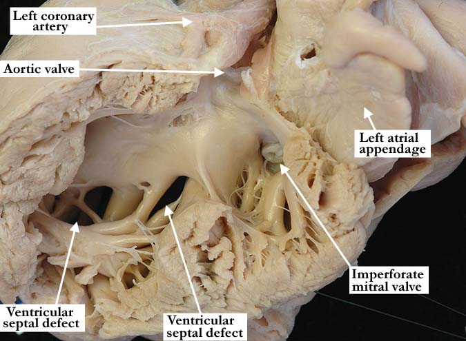
Modality: Anatomic specimen Orientation: Posterior view Description: In this view of the left ventricle there is an imperforate mitral valve within the inlet. It consists of a diaphragm-like valve with a single tendinous cord. The aortic valve guards the outlet. The two muscular ventricular septal defects are easily appreciated. Contributor: Diane E. Spicer, BS Institution: The Congenital Heart Institute of Florida (CHIF) Image Label: A060202-94a Source of Image: Van Mierop Archive, University of Florida Image Certification: pending AWG Rating: pending
|
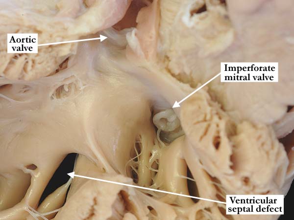
Modality: Anatomic specimen Orientation: Posterior view Description: A close-up view of the previous image demonstrates the imperforate, diaphragm-like mitral valve. Contributor: Diane E. Spicer, BS Institution: The Congenital Heart Institute of Florida (CHIF) Image Label: A060202-94b Source of Image: Van Mierop Archive, University of Florida Image Certification: pending AWG Rating: pending
|
|||
|
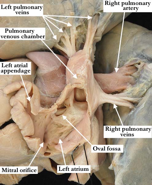
Modality: Anatomic specimen Orientation: Posterior lateral view Description: The left atrium and atrial appendages have been opened to demonstrate the left side of the atrial septum and oval fossa along with a hypoplastic mitral orifice. The left lung has been lifted away to view the pulmonary veins. There is a pulmonary component to the left atrium with all of the pulmonary veins draining to a common cavity superior to the main left atrial chamber. This is not entirely consistent with cor triatriatum because the constriction or ridge between the pulmonary venous chamber and the left atrium is not prominent enough. This would be considered a double-chambered left atrium. Contributor: Diane E. Spicer, BS Institution: The Congenital Heart Institute of Florida (CHIF) Image Label: A060202-94c Source of Image: Van Mierop Archive, University of Florida Image Certification: pending AWG Rating: pending
|
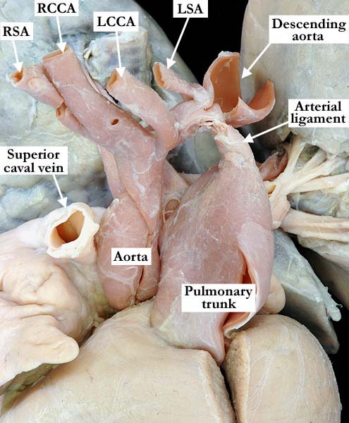
Modality: Anatomic specimen Orientation: Anterior view Description: The aorta and pulmonary trunk are normally related. The aorta is mildly hypoplastic, the brachiocephalic vessels branch normally from the arch and the arch extends to the left. There is tubular hypoplasia of the arch between the left common carotid (LCCA) and left subclavian (LSA) arteries. (RSA- right subclavian artery, RCCA- right common carotid artery) Contributor: Diane E. Spicer, BS Institution: The Congenital Heart Institute of Florida (CHIF) Image Label: A060202-94d Source of Image: Van Mierop Archive, University of Florida Image Certification: pending AWG Rating: pending |
|||
|
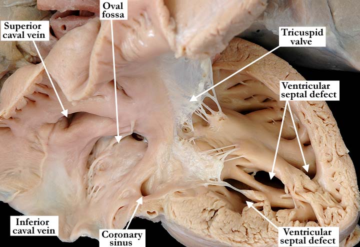
Modality: Anatomic specimen Orientation: Anterior view Description: In this anterior anatomic view, the free wall of the right atrium and ventricle have been lifted away. There are two muscular, ventricular septal defects, one of which is at the trabecular septum, extending toward the apex. The remaining ventricular septal defect is toward the inferior aspect of the trabecular septum. Contributor: Diane E. Spicer, BS Institution: The Congenital Heart Institute of Florida (CHIF) Image Label: A060202-94e Source of Image: Van Mierop Archive, University of Florida Image Certification: pending AWG Rating: pending
|
|
|||
AWG Page Certification: pending
|
Copyright ipccc-awg.net All Rights Reserved. Frontpage-Templates.org |