|
||||||||
|
|
|||||||||
|
|||||||||
|
IPCCC: 06.02.33 |
|||
|
AEPC Derived Term: |
Double orifice of mitral valve (06.02.33) |
||
|
EACTS-STS Derived Term: |
Mitral valve disease, Mitral valve pathology, Double orifice mitral valve (06.02.33) |
||
|
ICD 10 Term: |
Congenital malformation of aortic and mitral valves, unspecified (Q23.9) | ||
|
Comments: There are a series of subtypes of double orifice mitral valve listed as qualifiers in the IPCCC, such as the relative sizes of the two orifices and the position of the accessory (usually smaller) orifice. The images shown here are from different hearts with different subcategory attributes. Only the higher level codes are therefore used.
Definition: pending
|
|
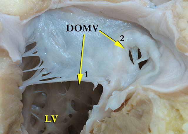
Modality: Anatomic specimen Orientation: Mitral valve view Description: This close up view of the mitral valve demonstrates a double orifice (DOMV). The smaller orifice (2) is suspended by several chordal attachments and has thickened edges adjacent to the lumen. The chordal attachments to the larger orifice (1) of the mitral valve that are on the same aspect as the smaller orifice are thickened and fused. (LV-left ventricle) Contributor: Diane Spicer, BS Institution: The Congenital Heart Institute of Florida (CHIF) Image Label: A060233-31a Source of Image: The Congenital Heart Institute of Florida (CHIF) Image Certification: 9 November 2013
AWG Rating:
|
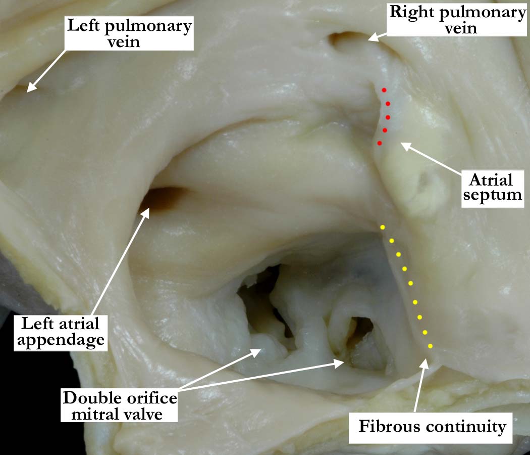
Modality: Anatomic specimen Orientation: Looking into the mitral valve from the opened left atrium at the base of the heart Description: From a different heart specimen and through the opened left atrium, the double orifice mitral valve is easily demonstrated. The smaller of the two orifices is closest to the interventricular septum and is in fibrous continuity with the aortic valve (yellow dots), while the larger orifice is in a more posterior position. The left atrium is dilated and the atrial septum is intact, the red dots indicating where the flap valve of the oval fossa is adherent to the left atrial endocardium. Contributor: Diane E. Spicer, BS Institution: The Congenital Heart Institute of Florida (CHIF) Image Label: A060233-31b Source of Image: Idriss Archive, Lurie Children's Hospital of Chicago, Chicago, IL, USA Image Certification: 9 November 2013
AWG Rating:
|
|||
|
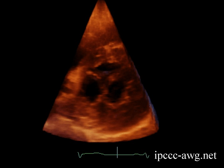
Modality: Transthoracic 3D echocardiogram Orientation: Short axis view Description: This is a three dimensional parasternal short axis view of a double orifice mitral valve obtained from another patient and seen from apex (looking up from the left ventricle). Contributor: Javier H. Gonzalez, MD Institution: The Congenital Heart Institute of Florida (CHIF) Image Label: E060233-31c Source of Image: Medical University of South Carolina. Charleston, South Carolina, USA Image Certification: 9 November 2013
AWG Rating:
|
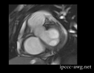
Modality: Cardiac MRI image (steady state free precession 2D cine image) Orientation: Short axis view Description: This image, from a different patient, illustrates a short axis view of a double orifice mitral valve. In this patient the superior valve orifice is larger than the inferior orifice. In combination, both valves function normally. Contributor: Marina Hughes, DPhil, MRCP, FRACP Institution: Centre for Cardiovascular Imaging, Great Ormond Street Hospital for Children NHS Trust, London, UK Image Label: MRA060233-31d Source of Image: Centre for Cardiovascular Imaging, Great Ormond Street Hospital for Children NHS Trust, London, UK Image Certification: 9 November 2013
AWG Rating:
|
|||
AWG Page Certification: 9 November 2013
|
Copyright ipccc-awg.net All Rights Reserved. Frontpage-Templates.org |