|
||||||||
|
|
|||||||||
|
|||||||||
|
IPCCC: 12.37.01, 15.95.30, 15.41.13, 10.09.14, 10.18.31
|
|||
|
AEPC Derived Term: |
Heart transplant (12.37.01) Post heart transplant graft rejection (15.95.30) Cardiac transplant associated coronary allograft vasculopathy (15.41.13) Acquired coronary occlusion (10.09.14) Old myocardial infarction (10.18.31) |
||
|
EACTS-STS Derived Term: |
Transplant, Heart (12.37.01) Coronary artery allograft vasculopathy (15.41.13) Coronary artery occlusion (10.09.14) Myocardial infarction, Old (10.18.31) |
||
|
ICD10 Derived Term: |
Heart transplant status (Z94.1) Heart transplant failure and rejection (T86.2) Coronary (artery): disease (I25.1) Other acute ischaemic heart diseases (I24) Old myocardial infarction (I25.2) |
||
|
Definition: pending
|
|
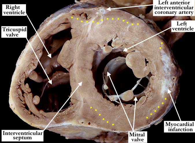
Modality: Anatomic specimen Orientation: Short axis - apical view Description: This simulated, short axis, echocardiographic view demonstrates the subendocardial (yellow dots) and mid septal myocardial congestion in this heart with rejection and ischemia. At the mid free wall there is a white scarred area consistent with an old myocardial infarct. The left anterior interventricular coronary artery was occluded at multiple sites along its length. Contributor: Diane E. Spicer, BS Institution: The Congenital Heart Institute of Florida (CHIF) Image Label: A123701-97a Source of Image: All Children's Hospital, St. Petersburg, Florida Image Certification: pending AWG Rating: pending
|
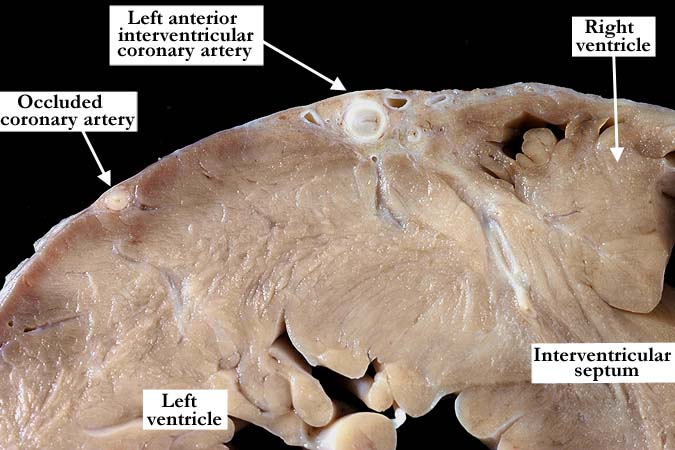
Modality: Anatomic specimen Orientation: Short axis view Description: This close-up view illustrates an occluded area along the length of the left anterior interventricular coronary artery. Note the small occluded artery just to the left. Contributor: Diane E. Spicer, BS Institution: The Congenital Heart Institute of Florida (CHIF) Image Label: A123701-97b Source of Image: All Children's Hospital, St. Petersburg, Florida Image Certification: pending AWG Rating: pending |
|||
|
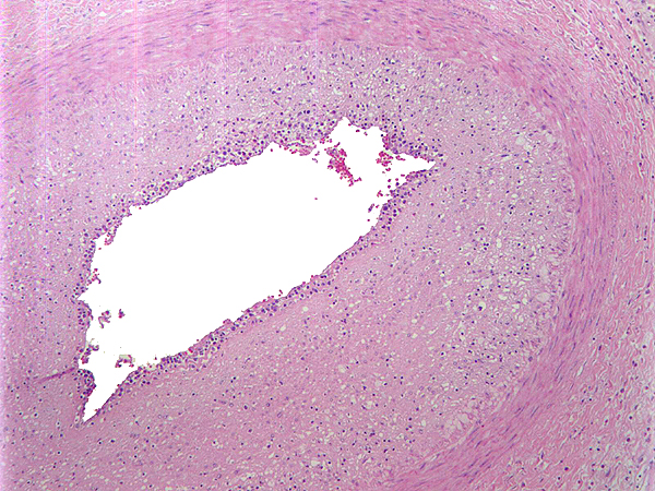
Modality: Microscopic section Orientation: High power Description: Cross section of an epicardial artery showing concentric intimal fibromyxoid and inflammatory thickening, narrowing the lumen by 70 %. There is subintimal lymphocytic and monocyte cell infiltrate, no mural arteritis or fibrinous necrosis is seen. Contributor: Hector Monforte, MD Institution: All Children's Hospital, St. Petersburg, Florida Image Label: MS123701-97c Source of Image: All Children's Hospital, St. Petersburg, Florida Image Certification: pending AWG Rating: pending
|
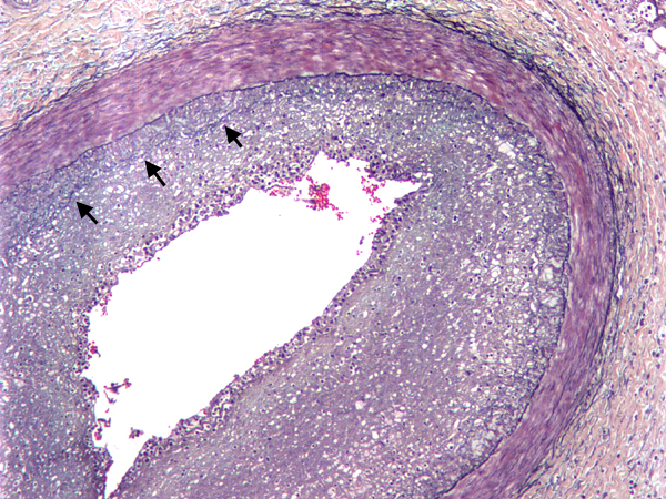
Modality: Microscopic section Orientation: High power, special stain Description: Movat pentachrome stain showing intact and preserved internal and external elastic laminae and muscular wall. The intimal thickening shows variable organization and inflammatory cells. Focally there is layering and duplication of the internal elastic lamina (arrows) Contributor: Hector Monforte, MD Institution: All Children's Hospital, St. Petersburg, Florida Image Label: MS123701-97d Source of Image: All Children's Hospital, St. Petersburg, Florida Image Certification: pending AWG Rating: pending
|
|||
AWG Page Certification: pending
|
Copyright ipccc-awg.net All Rights Reserved. Frontpage-Templates.org |