|
|||||
|
|
||||||
|
||||||
|
IPCCC: 01.01.09, 09.15.03, 06.02.92, 06.06.01, 06.07.05,
05.06.01, 04.01.02, 04.04.02, 04.01.03 |
|||
|
AEPC Derived Term: |
Hypoplastic left heart syndrome, (01.01.09) Aortic atresia (09.15.03) Mitral stenosis (06.02.92) (see Commentary below) AVSD: isolated atrial component (primum ASD)(partial) (06.06.01) AVSD with ventricular imbalance: dominant right ventricle, hypoplastic left ventricle, (06.07.05) Common atrium (virtual absence of atrial septum), (05.06.01) Left superior caval vein (SVC) persisting to left-sided atrium (04.01.02) Coronary sinus defect in left atrium: completely unroofed (04.04.02) Bilateral superior caval veins (SVC) (04.01.03) |
||
|
EACTS-STS Derived Term: |
Hypoplastic left heart syndrome (HLHS), Aortic atresia + Mitral stenosis (01.01.09, 09.15.03, 06.02.92) (see Commentary below) AVC (AVSD), Partial (incomplete) (PAVSD) (ASD primum with no VSD), Unbalanced, Small LV (06.06.01, 06.07.05) ASD, Common atrium (single atrium) (05.06.01) Systemic venous anomaly, SVC, Bilateral SVC, LSVC to left-sided atrium (completely unroofed CS) (04.01.02, 04.04.02, 04.01.03) |
||
|
Definition: pending
Commentary: Some authors would describe this as a stenotic left sided component of the AV valve not as 'Mitral' stenosis. |
|
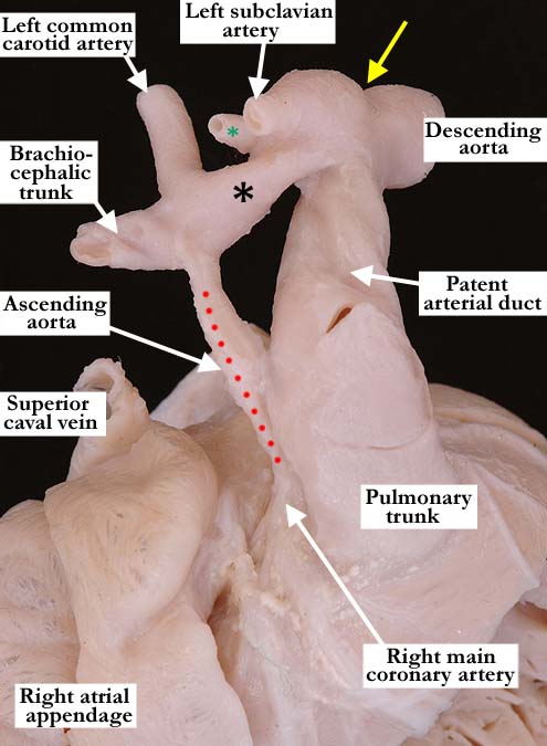
Modality: Anatomic specimen Orientation: Right, lateral, antero-superior view Description: This close up, anterior superior view of the great vessels in this heart with hypoplastic left heart illustrates the severe hypoplasia of the ascending aorta, a narrow aortic arch (black asterisk) and a juxtaductal constriction (yellow arrow) representing a coarctation. The brachiocephalic vessels branch normally from the aortic arch with a left vertebral artery (green asterisk) arising adjacent to the left subclavian artery. There is dilatation of the aorta proximal to the coarctation and note the direct extension of the patent arterial duct into the descending aorta. Contributor: Diane E. Spicer, BS Institution: The Congenital Heart Institute of Florida (CHIF) Image Label: A010109-83a Source of Image: Van Mierop Archive, University of Florida Image Certification: pending AWG Rating: pending
|
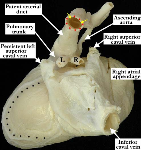
Modality: Anatomic specimen Orientation: Posterior view Description: The posterior view of the same heart shown in a, shows the large pulmonary trunk and arterial duct with the normal bifurcation of the right (R) and left (L) pulmonary arteries. The juxtaductal ridge (yellow arrows) can be seen through the opening of the descending aorta (red dots). There is a persistent left superior caval vein. The delimiting coronary arteries are marked by the black dots and outline the hypoplastic left ventricle on the posterior aspect of the heart. Contributor: Diane E. Spicer, BS Institution: The Congenital Heart Institute of Florida (CHIF) Image Label: A010109-83b Source of Image: Van Mierop Archive, University of Florida Image Certification: pending AWG Rating: pending |
|||
|
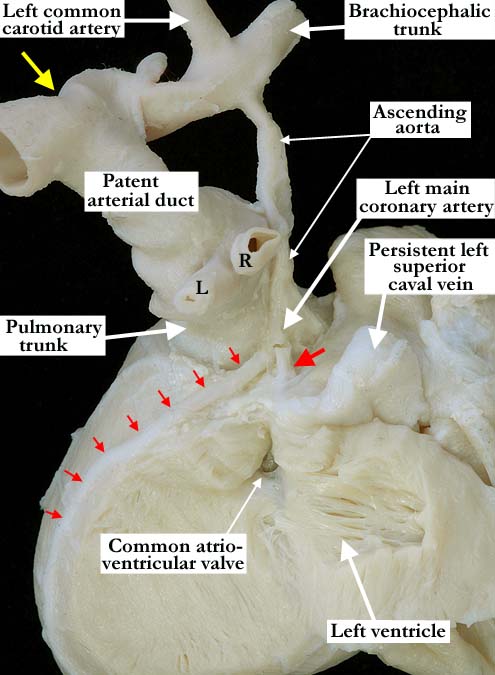
Modality: Anatomic specimen Orientation: Posterior view Description: The hypoplastic left ventricle is slit-like without evident endocardial fibroelastosis, the majority of the chamber left attached to the free wall. There is an unbalanced, atrioventricular septal defect of the primum type. The left anterior interventricular coronary artery (small red arrows) and the circumflex coronary artery (single large red arrow) branch normally from the left main coronary artery. The large, patent arterial duct demonstrates the characteristic, direct extension to the descending aorta with the yellow arrow marking the juxtaductal coarctation. Contributor: Diane E. Spicer, BS Institution: The Congenital Heart Institute of Florida (CHIF) Image Label: A010109-83c Source of Image: Van Mierop Archive, University of Florida Image Certification: pending AWG Rating: pending
|
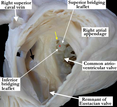
Modality: Anatomic specimen Orientation: Right lateral view Description: This close up, anterior lateral view through the right atrial appendage demonstrates the common atrium in this heart with a severely unbalanced atrioventricular septal defect and hypoplastic left heart. The superior caval vein enters the right atrium in the usual fashion. The black arrow represents the entry of the persistent left superior caval vein into the common atrium, with absence of the roof of the coronary sinus. A thin remnant of the Eustachian valve extends along the inferior wall of this common atrium. The tiny, left component of the unbalanced atrioventricular septal defect is marked by the yellow arrow. The superior and inferior bridging leaflets are fused to one another (red dot) and to the crest of the ventricular septum. Contributor: Diane E. Spicer, BS Institution: The Congenital Heart Institute of Florida (CHIF) Image Label: A010109-83d Source of Image: Van Mierop Archive, University of Florida Image Certification: pending AWG Rating: pending
|
|||
AWG Page Certification: pending
|
Copyright ipccc-awg.net All Rights Reserved. Frontpage-Templates.org |