|
|||||
|
|
||||||
|
||||||
|
IPCCC: 01.01.04, 06.02.01, 07.11.02, 05.04.07, 07.07.00, 12.14.02
or 01.01.22, 06.02.01, 01.01.04 vs 01.01.19 |
|||
|
AEPC Derived Term: |
Double outlet right ventricle (01.01.04) Mitral atresia (06.02.01) Muscular VSD in inlet septum (07.11.02) Atrial septal defect (ASD) within oval fossa (secundum): fenestrated (multiple defects) (05.04.07) Left ventricular hypoplasia (07.07.00) Pulmonary trunk band (PA band) (12.14.02) |
||
|
EACTS-STS Derived Term: |
Single ventricle, Mitral atresia, DORV (01.01.22, 06.02.01, 01.01.04) or DORV, Remote VSD (Uncommitted VSD)(01.01.19) Mitral atresia (06.02.01) plus ASD, Secundum, Fenestrated, Multiple holes (05.04.07) Hypoplastic left ventricle (07.07.00) PA banding (12.14.02) |
||
|
Definition: pending |
|
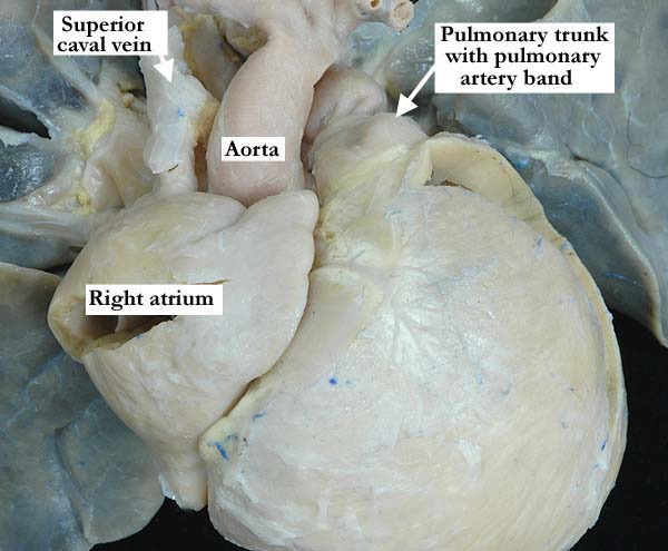
Modality: Anatomic specimen Orientation: Anterior view Description: This view demonstrates the superior caval vein entering the right sided atrium that on outward examination is consistent with a morphologically right atrial appendage. The aorta and pulmonary trunk appear normally related, with a pulmonary artery band in place. Contributor: Diane Spicer, BS Institution: The Congenital Heart Institute of Florida (CHIF) Image Label: A010104-72a Source of Image: Idriss Archive, Childrens Memorial Hospital, Chicago, IL Image Certification: pending AWG Rating: pending
|
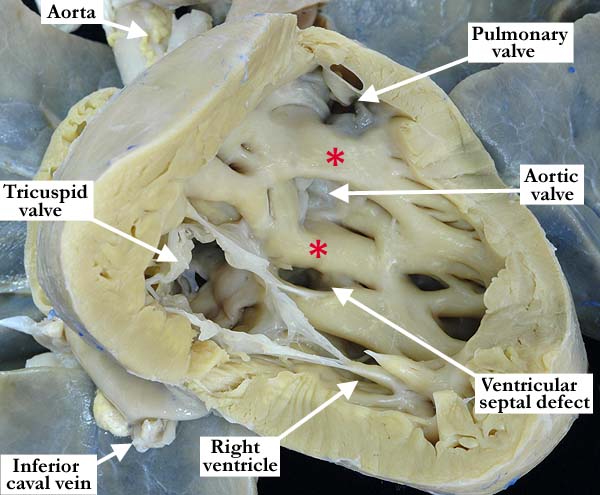
Modality: Anatomic specimen Orientation: Anterior view of the right ventricle Description: The right ventricle has been opened in a clam shell-like fashion, showing the tricuspid valve guarding the inlet and the origin of both arterial trunks from this double outlet right ventricle. There are bilateral, complete, muscular infundibulums (red asterisks) supporting each arterial trunk with absence of continuity between the leaflets of the arterial and atrioventricular valves. There is a non-committed, restrictive, muscular, inlet ventricular septal defect. Note the coarse trabeculations within this morphologically right ventricle. Contributor: Diane Spicer, BS Institution: The Congenital Heart Institute of Florida (CHIF) Image Label: A010104-72b Source of Image: Idriss Archive, Childrens Memorial Hospital, Chicago, IL Image Certification: pending AWG Rating: pending
|
|||
|
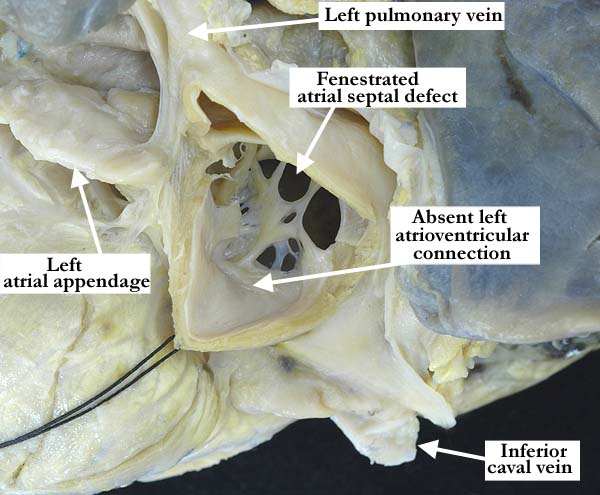
Modality: Anatomic specimen Orientation: Left atrial view Description: This view into the left atrium of the heart shown above, illustrates a fenestrated atrial septal defect and absence of the left atrioventricular connection. Contributor: Diane Spicer, BS Institution: The Congenital Heart Institute of Florida (CHIF) Image Label: A010104-72c Source of Image: Idriss Archive, Childrens Memorial Hospital, Chicago, IL Image Certification: pending AWG Rating: pending
|
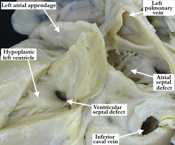
Modality: Anatomic specimen Orientation: Posterior view Description: This posterior view of the heart shown above, illustrates a hypoplastic, morphologically left ventricle with its fine apical trabeculations. The restrictive muscular ventricular septal defect lies in the inlet portion of the interventricular septum in this heart with double outlet right ventricle and absent left atrioventricular connection. Contributor: Diane Spicer, BS Institution: The Congenital Heart Institute of Florida (CHIF) Image Label: A010104-72d Source of Image: Idriss Archive, Childrens Memorial Hospital, Chicago, IL Image Certification: pending AWG Rating: pending
|
|||
AWG Page Certification: pending
|
Copyright ipccc-awg.net All Rights Reserved. Frontpage-Templates.org |