Images & Codes 


Archiving Working Group

International Society for Nomenclature of

Paediatric and Congenital Heart Disease

ipccc-awg.net


               Close Window    

IPCCC: 01.01.22, 06.02.01, 01.01.04, 06.02.02, 01.04.00,

[07.10.00], 07.10.17, 07.11.07, 02.06.02, 07.07.00, 10.10.12


AEPC Derived Term:    

Functionally 
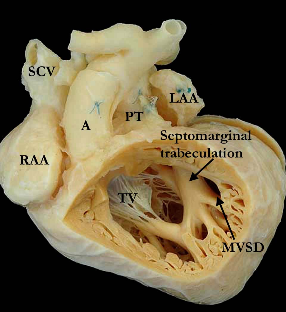
univentricular heart (01.01.22)
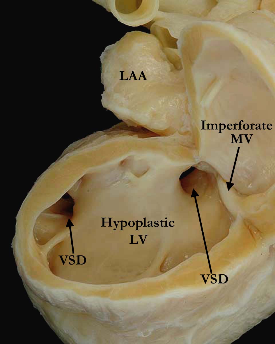
Mitral atresia (06.02.01)
Double outlet right ventricle (01.01.04)
Mitral valve atretic (imperforate) (06.02.02)
Concordant atrioventricular connections (01.04.00)
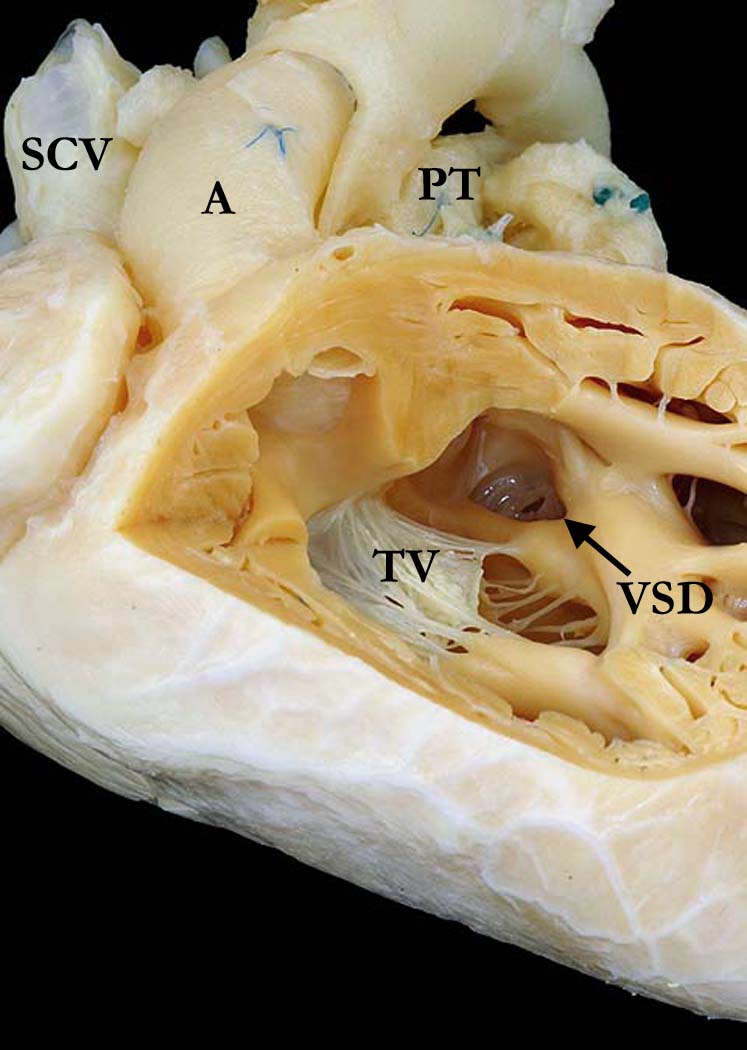
Ventricular septal defect (VSD) + malaligned outlet septum anteriorly (07.10.17)
Muscular ventricular septal defect (VSD) in anterior septum (07.11.07)
Aortic orifice anterior right with respect to pulmonary orifice (02.06.02)
Left ventricular hypoplasia (07.07.00)

Endocardial fibroelastosis (10.10.12)


EACTS-STS Derived Term:    

Single ventricle, Mitral atresia, DORV (01.01.22, 06.02.01, 01.01.04)
Mitral valve disease, Mitral valve pathology, Mitral valve atretic (imperforate)(06.02.02)
AV connection(s) = Normal atrioventricular connections (Concordant atrioventricular connections in biventricular heart) (01.04.00)
VSD-modifier for infundibular septal morphology, VSD + malaligned outlet septum, Anterior deviation of infundibular septum (07.10.00, 07.10.17)
VSD, Type 4 (Muscular), Trabecular, Anterior (07.11.07)
Relationship of aortic orifice with respect to pulmonary orifice, Aortic orifice right and anterior with respect to pulmonary orifice (02.06.02)
DORV-modifier, Hypoplastic left ventricle (07.07.00)
Endocardium disease[s], Endocardial fibroelastosis (10.10.12)


ICD10 Derived Term:  

Congenital malformation of cardiac chambers and connections, unspecified (Q20.9)
Double outlet right ventricle (Q20.1)
Congenital mitral stenosis: Congenital mitral atresia (Q23.2)
Ventricular septal defect (Q21.0)
Other congenital malformations of cardiac chambers and connections (Q20.8)
Endocardial fibroelastosis (I42.4)


Definition:  NA
 

Commentary: NA


Modality: Anatomic specimen

Orientation: Right ventricular view

Description:  This figure shows one of two views of the same right ventricle in this heart with imperforate mitral valve, left ventricular hypoplasia and double outlet right ventricle. The aorta (A) is anterior and to the right of the pulmonary trunk (PT). The trabeculations within the right ventricle are prominent with a freestanding septomarginal trabeculation. On the anterosuperior aspect of the ventricular septum and anterior to the septomarginal trabeculation, there is the opening of a muscular ventricular septal defect (MVSD) better seen in the lower panel (Image: A010104-29c). The close up view on the right sided panel (Image: A010104-29b), shows the right ventricular apex tilted upwards so that the double outlet is easily visualized, along with the malaligned, subpulmonary ventricular septal defect (VSD) through which the imperforate mitral valve is seen. The ventricular septal defect has a posterior inferior muscular rim separating it from the tricuspid valve (TV). (SCV - superior caval vein, RAA - right atrial appendage, LAA - left atrial appendage, A - aorta, PT - pulmonary trunk).

Contributor: Diane Spicer, BS

Institution: The Congenital Heart Institute of Florida (CHIF)

Image Label:   A010104-29a

Source of Image: Van Mierop Archive, University of Florida, Gainesville, FL

Image Certification: 13 July 2013

AWG Rating


Modality: Anatomic specimen

Orientation:  Right ventricular view

DescriptionThis is the companion view of the left sided panel (Image: A010104-29a).

Contributor: Diane Spicer, BS

Institution: The Congenital Heart Institute of Florida (CHIF)

Image Label:    A010104-29b

Source of Image:  Van Mierop Archive, University of Florida, Gainesville, FL

Image Certification: 13 July 2013

AWG Rating


Modality: Anatomic specimen

Orientation: Left ventricular view

Description:  The left ventricular view of the heart shown in image one shows a hypoplastic left ventricle with endocardial fibroelastosis. The mitral valve is imperforate. The two ventricular septal defects (VSD) are easily appreciated, one anterior and one at the base of the heart (malaligned). (LAA - left atrial appendage).

Contributor: Diane Spicer, BS

Institution: The Congenital Heart Institute of Florida (CHIF)

Image Label:  A010104-29c

Source of Image: Van Mierop Archive, University of Florida, Gainesville, FL

Image Certification: 13 July 2013

AWG Rating


AWG Page Certification: 13 July 2013

Copyright ipccc-awg.net   All Rights Reserved. Frontpage-Templates.org