|
|||||
|
|
||||||
|
||||||
|
IPCCC Code:
09.45.00, 01.01.07 |
|||
|
AEPC Code: |
Coronary fistula(s)/ sinusoids, Pulmonary atresia + intact ventricular septum | ||
|
EACTS-STS Code: |
Pulmonary atresia-IVS-modifier-Coronary artery fistula(s) or sinusoid(s), (Coronary-cameral fistula(s)) present | ||
|
Definition: NA
|
|
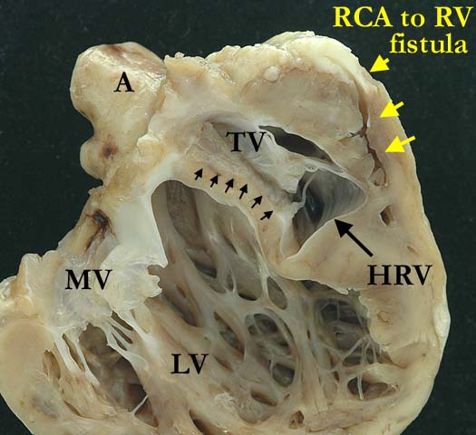
This four chamber view of a heart with pulmonary atresia and intact ventricular septum demonstrates a hypoplastic right ventricle (HRV) with a right coronary artery (RCA) to right ventricular (RV) fistula. The right coronary artery is dilated and thick walled along nearly its entire length. The interventricular septum is thin and there is an Ebstein-like malformation of the septal leaflet of the tricuspid valve (TV). The septal leaflet is adherent to the septum along is entire length (black arrows). (A-aorta, LV-left ventricle, MV-mitral valve)
Contributor: Diane Spicer, BS Image Name: CAanom1.jpg
|
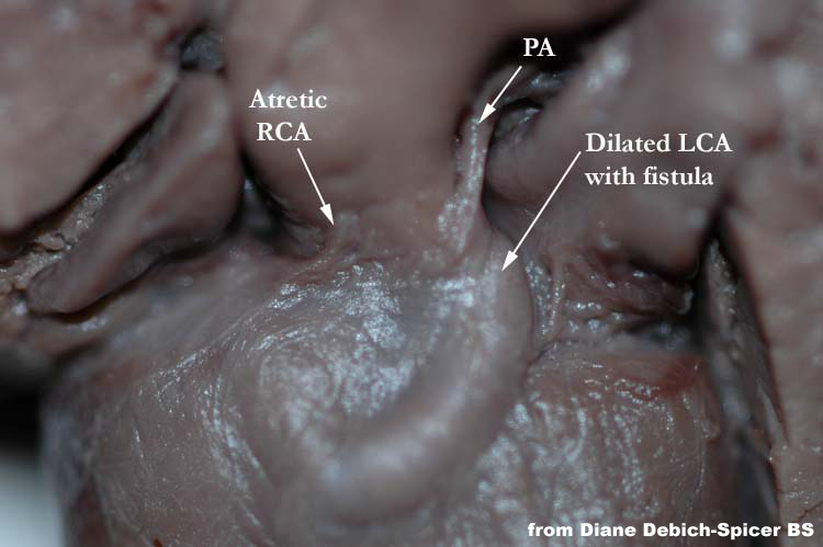
This superior view of a heart with pulmonary atresia and intact ventricular septum (not shown) demonstrates an atretic pulmonary artery (PA) with a left coronary artery (LCA) to right ventricular fistula (not shown). The left coronary artery is dilated along nearly its entire length.
Contributor: Diane Spicer, BS Image Name: PAA_RCA_Atresia.jpg |
|||
|
|
||||
|
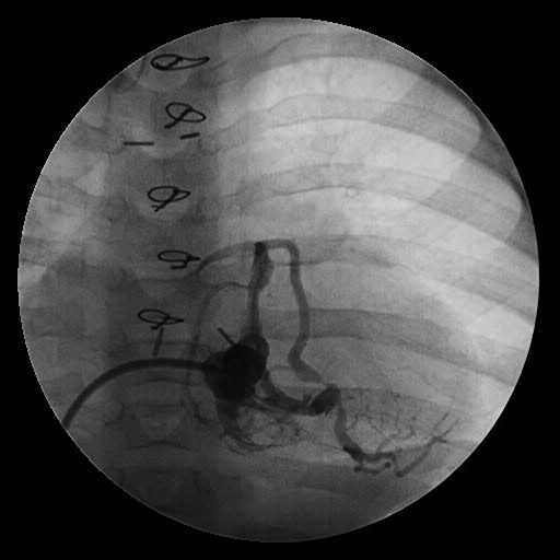
This angiocardiogram from a patient with pulmonary atresia and intact ventricular septum, illustrates the fistulous connection between the left coronary artery and the hypoplastic right ventricular cavity. Note that there is a significant obstruction in the mid-portion of the anterior descending coronary artery created by the fistulous connection. Flow occurs only during systole and there is retrograde filling of the ascending aorta.
Contributor: Jorge M. Giroud, MD
|
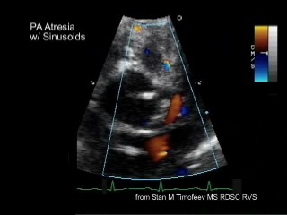
This echocardiogram from a patient with pulmonary atresia illustrates the fistulous connection between the left coronary artery and the hypoplastic right ventricular cavity. Note that the color doppler evaluation demonstrates the abnormal connections between the coronary arteries and right ventricle but cannot image obstructions to the coronary arteries with the precision of an angiogram.
Contributor: Stan Timofeev, MS
|
|||
AWG Certification: Pending
|
Copyright ipccc-awg.net All Rights Reserved. Frontpage-Templates.org |