|
||||||||
|
|
|||||||||
|
|||||||||
|
IPCCC Code: 09.29.02 |
|||
|
AEPC Derived Term: |
Preductal aortic coarctation (09.29.02) | ||
|
EACTS-STS Derived Term: |
Coarctation of the Aorta (CoAo)-modifier, Preductal (Infantile type) (09.29.02) | ||
|
ICD10 Derived Term: |
Coarctation of aorta (Q25.1) | ||
|
Definition: NA |
|
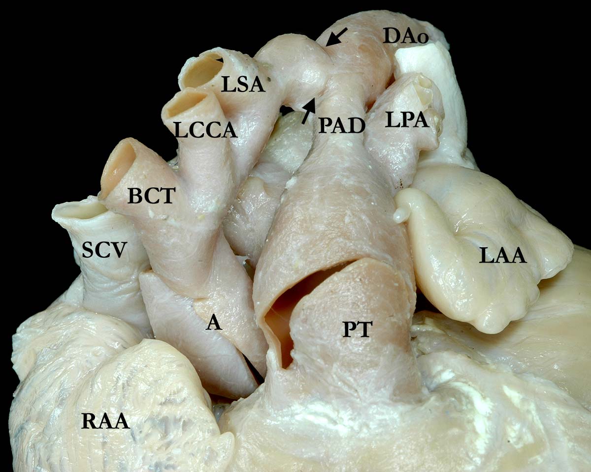
Modality: Anatomic specimen Orientation: Anterior view of the base of the heart Description: The great arteries exit the ventricular mass in the normal fashion in this anatomic view of the base of the heart. The brachiocephalic vessels branch from the aortic arch in the usual fashion, the ascending aorta and the aortic arch smaller than normal. Just distal to the left subclavian artery (LSA) and proximal to where the patent arterial duct (PAD) joins the descending aorta (DAo), there is a coarctation (black arrows). There is some of post stenotic dilatation of the aorta. (SCV-superior caval vein, RAA-right atrial appendage, LAA-left atrial appendage, PT-pulmonary trunk, LPA-left pulmonary artery, BCT-brachiocephalic trunk, LCCA-left carotid artery) Contributor: Diane Spicer, BS Institution: Congenital Heart Institute of Florida Image Label: A092902-52a Source of Image: Van Mierop Archive, University of Florida, Gainesville, Florida Image Certification: pending AWG Rating: pending |
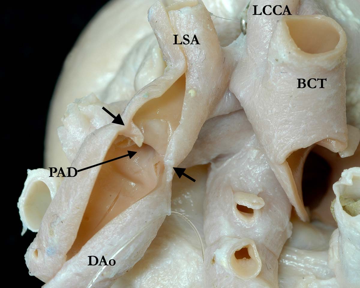
Modality: Anatomic specimen Orientation: Posterior view of the base of the heart Description: The great arteries exit the ventricular mass in the normal fashion in this anatomic view of the base of the heart. The brachiocephalic vessels branch from the aortic arch in the usual fashion, the ascending aorta and the aortic arch smaller than normal. Just distal to the left subclavian artery (LSA) and proximal to where the patent arterial duct (PAD) joins the descending aorta (DAo), there is a coarctation (black arrows). There is some of dilatation of the descending aorta, distal to the coarctation. (SCV-superior caval vein, RAA-right atrial appendage, LAA-left atrial appendage, PT-pulmonary trunk, LPA-left pulmonary artery, BCT-brachiocephalic trunk, LCCA-left carotid artery) Contributor: Diane Spicer, BS Institution: Congenital Heart Institute of Florida Image Label: A092902-52b Source of Image: Van Mierop Archive, University of Florida, Gainesville, Florida Image Certification: pending AWG Rating: pending
|
|||
|
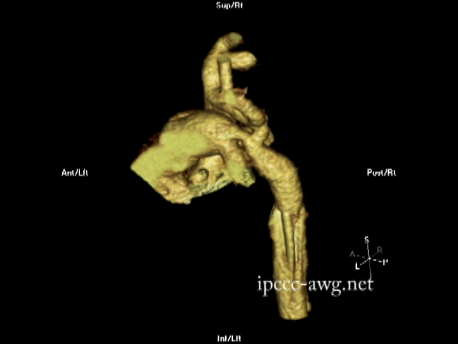
Modality: CT Angiogram Orientation: Three dimensional video Description: 3 dimensional reconstruction of a CT angiographic image. Isolated coarctation of the aorta just distal to the left subclavian artery, in the juxta-arterial duct postion. There is post stenotic dilation of the proximal descending aorta. An umbilical arterial catheter causes artifact in the descending thoracic aorta Contributor: Charles W. Shepard, MD Institution: University of Minnesota Amplatz Childrens Hospital Image Label: A092902-52c Image Source: Heart Center, University of Minnesota Amplatz Childrens Hospital Minneapolis, Minnesota Image Certification: pending AWG Rating: pending |
|
|||
AWG Certification: Pending
|
Copyright ipccc-awg.net All Rights Reserved. Frontpage-Templates.org |