|
||||||||
|
|
|||||||||
|
|||||||||
|
IPCCC: 10.03.28 |
|||
|
AEPC Derived Term: |
Benign heart tumour - multiple rhabdomyoma (10.03.28) |
||
|
EACTS-STS Derived Term: |
Cardiac tumor, Primary, Rhabdomyoma, Multiple (10.03.28) |
||
|
ICD 10 Term: |
Benign neoplasm of other and unspecified intrathoracic organs, Heart (D15.1) | ||
|
Definition: pending
|
|
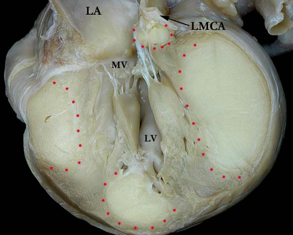
Modality: Anatomic specimen Orientation: Longitudinal section
Description: This cross section of the
left ventricle shows multiple rhabdomyomas within the myocardium (red dots).
Rhabdomyomas are commonly associated with tuberous sclerosis. The left
coronary artery (LMCA)
can be seen at the atrioventricular junction with a mass just inferior to
it. The location of the mass has the potential to compress the coronary
artery. (LA - left atrium,
MV - mitral valve,
LV - left ventricle) (Reference: Geva T, Santini F, Pear W, Driscoll SG, Van Praagh R. Cardiac rhabdomyoma.
Rare cause of fetal death. Chest.1991;99(1):139-142. ) Institution: The Congenital Heart Institute of Florida (CHIF) Image Label: A100328-25a Source of Image: The Congenital Heart Institute of Florida (CHIF) Image Certification: 8 September 2012
AWG Rating:
|
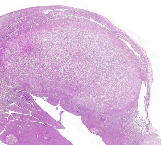
Modality: Photomicrograph Description: Low power H&E photomicrograph of a cross section of the left ventricle with a conspicuous 2cm rhabdomyoma and several incipient microscopic nodular lesions. Contributor: Hector L. Monforte, MD Institution: The Congenital Heart Institute of Florida (CHIF) Image Label: P100328-25b Source of Image: All Children's Hospital, St. Petersburg, Florida Image Certification: 8 September 2012
AWG Rating:
|
|||
|
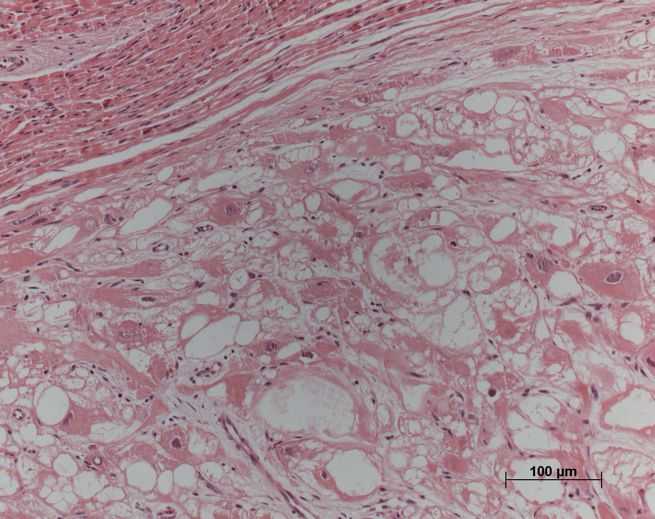
Modality: Photomicrograph Description: High-power-field protomicrograph of a cardiac rhabdomyoma in a different patient, showing the enlarged and vacuolated neoplastic cells, some presenting the typical "spider-cell" morphology. For reference, at the left upper corner there is the adjacent normal myocardium. Hematoxylin-eosin stain. The bar at the right lower corner represents 100 micrometers. Contributor: Vera D. Aiello, MD Institution: Heart Institute (InCor), University of São Paulo Medical School, São Paulo, Brazil Image Label: P100328-25c Source of Image: Heart Institute (InCor), University of São Paulo Medical School, São Paulo, Brazil Image Certification: 8 September 2012
AWG Rating:
|
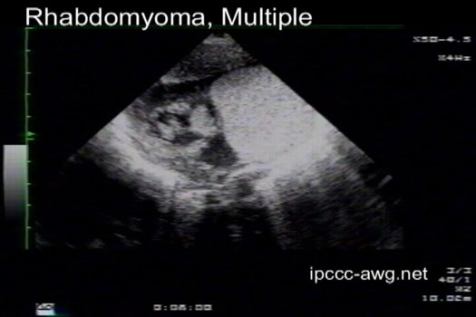
Modality: Echocardiogram Orientation: Multiple views Description: This video clip is from the same patient as shown in images 1 & 2 above. Note the tumors that dwarf the ventricular cavity. Contributor: Jorge M. Giroud, MD Institution: The Congenital Heart Institute of Florida (CHIF) Image Label: E100328-25d Source of Image: The Congenital Heart Institute of Florida (CHIF) Image Certification: 8 September 2012
AWG Rating:
|
|||
AWG Page Certification: 8 September 2012
|
Copyright ipccc-awg.net All Rights Reserved. Frontpage-Templates.org |