|
||||||||
|
|
|||||||||
|
|||||||||
|
IPCCC: 03.01.04, 01.03.02, 01.05.01, 04.06.00, 04.01.03, 04.04.09,
03.03.03, 06.06.00, 05.04.02, 09.46.22 |
|||
|
AEPC Derived Term: |
Right isomerism ('asplenia') (03.01.04) Isomerism of right atrial appendages (right isomerism) (01.03.02) Discordant VA connections (TGA) (01.05.01) Totally anomalous pulmonary venous connection - supracardiac (04.06.00) Bilateral superior caval veins (SVC) (04.01.03) Coronary sinus absent (04.04.09) Bilateral right bronchi (short - right isomerism) (03.03.03) Atrioventricular septal defect (06.06.00) Atrial septal defect (ASD) within oval fossa (secundum) (05.04.02) Abnormal coronary origin: circumflex from right coronary aortic sinus (09.46.22) |
||
|
EACTS-STS Derived Term: |
Atrial appendage isomerism, Right (03.01.04) Syndrome, Heterotaxy (heterotaxy syndrome) (visceral heterotaxy)-modifier, Isomerism, Isomerism of the right atrial appendages (01.03.02) Syndrome, Heterotaxy (heterotaxy syndrome) (visceral heterotaxy)-modifier, Asplenia syndrome VA connection =Discordant VA connections (TGA) (01.05.01) Total anomalous pulmonary venous connection (TAPVC), Type 1 (supracardiac) (04.06.00) Systemic venous anomaly, SVC, Bilateral SVC (04.01.03) Systemic venous anomaly,-non-Caval vein, Coronary sinus, Coronary sinus absent (04.04.09) Syndrome, Heterotaxy (heterotaxy syndrome) (visceral heterotaxy)-modifier, Bilateral right bronchi (short - right isomerism) AVC (AVSD) (06.06.00) ASD, Secundum (05.04.02) Coronary anomaly, AAOC (Anomalous aortic origin of coronary), Cx from RASV (right aortic sinus of Valsalva) |
||
|
Definition: pending
|
|
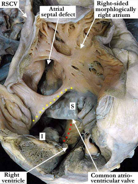
Modality: Anatomic specimen Orientation: Superior lateral view Description: This superior lateral view of the right-sided, morphologically right atrium and right ventricle, demonstrates an atrioventricular septal defect along with an atrial septal defect of the secundum type. The atrial border of the atrioventricular septal defect is marked with yellow dots and the crest of the interventriclar septum with red dots. The superior (S) and inferior (I) bridging leaflets extend over the crest of the ventricular septum. The right superior caval vein enters the roof of the right-sided, morphologically right atrium and the pectinate muscles encircle the atrioventricular junction. Contributor: Diane E. Spicer, BS Institution: The Congenital Heart Institute of Florida (CHIF) Image Label: A030104-100a Source of Image: Idriss Archive, Childrens Memorial Hospital, Chicago, IL Image Certification: pending AWG Rating: pending
|
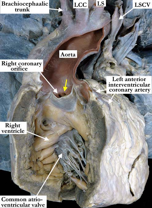
Modality: Anatomic specimen Orientation: Anterior view Description: The aorta arises from the opened right ventricular outflow tract. The brachiocephalic arteries branch from the aortic arch in the usual fashion and the arch extends to the left. The yellow arrow marks the coronary orifice that gives rise to the circumflex coronary artery. It is within the right aortic sinus and adjacent to the right coronary orifice. The common atrioventricular valve lies in the inlet and the right ventricle is hypertrophied. Contributor: Diane E. Spicer, BS Institution: The Congenital Heart Institute of Florida (CHIF) Image Label: A030104-100b Source of Image: Idriss Archive, Childrens Memorial Hospital, Chicago, IL Image Certification: pending AWG Rating: pending |
|||
|
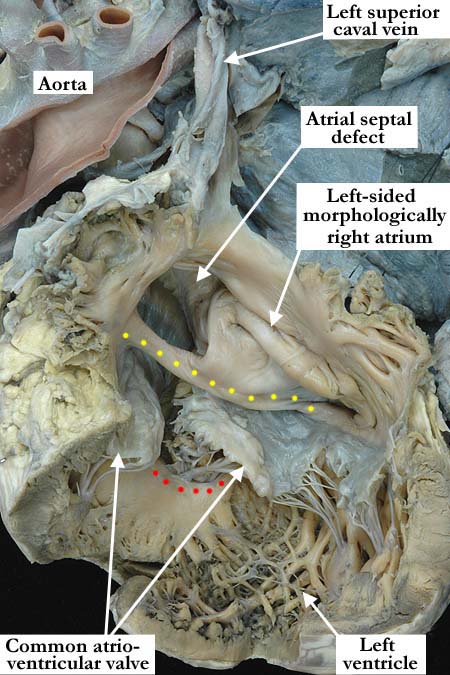
Modality: Anatomic specimen Orientation: Posterior view Description: The left-sided, morphologically right atrium and the left ventricle have been opened to demonstrate the common atrioventricular valve. The yellow dots mark the atrial septal border of the atrioventricular septal defect and the red dots are along the crest of the ventricular septum. There is a secundum type of atrial septal defect and a left superior caval vein drains directly into the roof of the left-sided, morphologically right atrium. The coronary sinus is absent. Note the pectinate muscles extending around the atrioventricular junction to the crux of the heart. Contributor: Diane E. Spicer, BS Institution: The Congenital Heart Institute of Florida (CHIF) Image Label: A030104-100c Source of Image: Idriss Archive, Childrens Memorial Hospital, Chicago, IL Image Certification: pending AWG Rating: pending
|
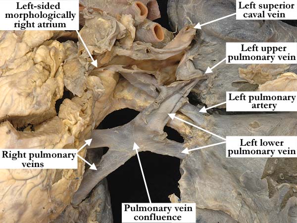
Modality: Anatomic specimen Orientation: Posterior view Description: The heart has been lifted toward the right and away from the pulmonary venous component. The right pulmonary veins and the left lower pulmonary vein join in a venous confluence that drains into the left superior caval vein. The left upper pulmonary vein enters the vertical vein just as it joins the left superior caval vein. Contributor: Diane E. Spicer, BS Institution: The Congenital Heart Institute of Florida (CHIF) Image Label: A030104-100d Source of Image: Idriss Archive, Childrens Memorial Hospital, Chicago, IL Image Certification: pending AWG Rating: pending
|
|||
|
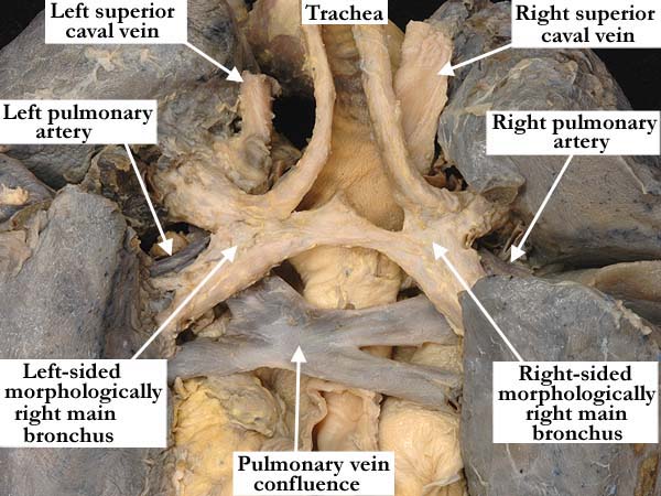
Modality: Anatomic specimen Orientation: Posterior view Description: This posterior view demonstrates bilateral, morphologically right bronchi. The bronchi are eparterial. The right and left superior caval veins lie on either side of the trachea and the pulmonary vein confluence is inferior to the carina. Contributor: Diane E. Spicer, BS Institution: The Congenital Heart Institute of Florida (CHIF) Image Label: A030104-100e Source of Image: Idriss Archive, Childrens Memorial Hospital, Chicago, IL Image Certification: pending AWG Rating: pending
|
|
|||
AWG Page Certification: pending
|
Copyright ipccc-awg.net All Rights Reserved. Frontpage-Templates.org |