|
||||||||
|
|
|||||||||
|
|||||||||
|
IPCCC: 03.01.05 [or 01.03.03], 04.01.03, 04.01.26, 04.03.02, 03.03.04,
06.06.00, 09.07.12, 12.05.01 |
|||
|
AEPC Derived Term: |
Left isomerism ('polysplenia') (03.01.05) Isomerism of left atrial appendages (left isomerism) (01.03.03) Bilateral superior caval veins (SVC) (04.01.03) Left superior caval vein (SVC) persisting to coronary sinus to right-sided atrium (04.01.26) Right-sided azygos continuation of inferior caval vein (IVC) (absent suprarenal segment) to right superior caval vein (SVC) (04.03.02) Bilateral left bronchi (long - left isomerism) (03.03.04) Atrioventricular septal defect (06.06.00) Pulmonary trunk dilation (09.07.12) AVSD: complete (common valve orifice) repair (12.05.01) |
||
|
EACTS-STS Derived Term: |
Atrial appendage isomerism, Left (03.01.05) Syndrome, Heterotaxy (heterotaxy syndrome) (visceral heterotaxy)-modifier, Polysplenia syndrome Systemic venous anomaly, SVC, Bilateral SVC, LSVC to CS (intact) to right-sided atrium (04.01.03, 04.01.26) Systemic venous anomaly, IVC, Abnormal RIVC, Interrupted RIVC, Azygos continuation (absent suprarenal segment) to RSVC (04.03.15, 04.03.02) Syndrome, Heterotaxy (heterotaxy syndrome) (visceral heterotaxy)-modifier, Bilateral left bronchi (long - left isomerism) AVC (AVSD) (06.06.00) Pulmonary trunk dilation (09.07.12 ) AVC (AVSD) repair, Complete (CAVSD) (12.05.01) |
||
|
Definition: pending
|
|
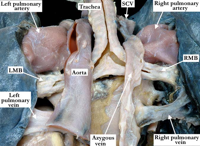
Modality: Anatomic specimen Orientation: Posterior view Description: This posterior view demonstrates a normal left aortic arch and descending aorta with a large, azygous vein arising from the superior caval vein. There is absence of the hepatic portion of the inferior caval vein with azygous vein continuation, hence its larger than normal size. The right and left pulmonary arteries are markedly dilated. The right (RMB) and left (LMB) main bronchi are bilaterally long and consistent with morphologically left main bronchi. Contributor: Diane E. Spicer, BS Institution: The Congenital Heart Institute of Florida (CHIF) Image Label: A030105-99a Source of Image: Idriss Archive, Childrens Memorial Hospital, Chicago, IL Image Certification: pending AWG Rating: pending
|
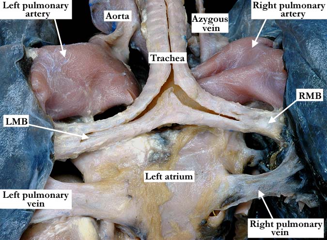
Modality: Anatomic specimen Orientation: Posterior view Description: The azygous vein and the aorta have been lifted away to demonstrate the bilateral, long, morphologically left main bronchi. The right and left pulmonary arteries are markedly dilated. (RMB-right main bronchus, LMB-left main bronchus) Contributor: Diane E. Spicer, BS Institution: The Congenital Heart Institute of Florida (CHIF) Image Label: A030105-99b Source of Image: Idriss Archive, Childrens Memorial Hospital, Chicago, IL Image Certification: pending AWG Rating: pending |
|||
|
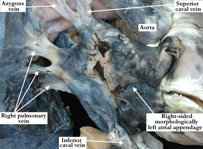
Modality: Anatomic specimen Orientation: Anterior lateral view Description: This anterior lateral view shows the external appearance of the right-sided, morphologically left atrial appendage. Although the outward appearance of this atrial appendage may not be entirely consistent with a morphologically left atrial appendage, the true arbiture will be the extent of the pectinate muscles. There are multiple sutures present, secondary to previous surgical intervention. Contributor: Diane E. Spicer, BS Institution: The Congenital Heart Institute of Florida (CHIF) Image Label: A030105-99c Source of Image: Idriss Archive, Childrens Memorial Hospital, Chicago, IL Image Certification: pending AWG Rating: pending
|
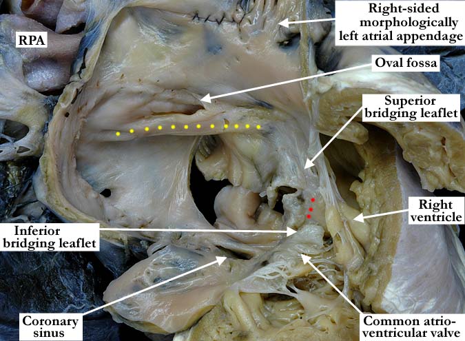
Modality: Anatomic specimen Orientation: Anterior oblique view Description: The right-sided atrium and atrial appendage, along with the right ventricle have been opened in a clam-shell fashion demonstrating the smooth atrial vestibule consistent with a morphologically left atrium. The upper portion of the atrial septum is well formed with a slit-like oval foramen. The yellow dots illustrate the atrial border of the atrioventricular septal defect. The red dots are along the crest of the ventricular septum. Above the oval foramen and extending around the inside of the atrium, to the coronary sinus, there are multiple small holes from a surgical patch that had been removed to show the atrioventricular septal defect. (RPA-right pulmonary artery) Contributor: Diane E. Spicer, BS Institution: The Congenital Heart Institute of Florida (CHIF) Image Label: A030105-99d Source of Image: Idriss Archive, Childrens Memorial Hospital, Chicago, IL Image Certification: pending AWG Rating: pending
|
|||
|
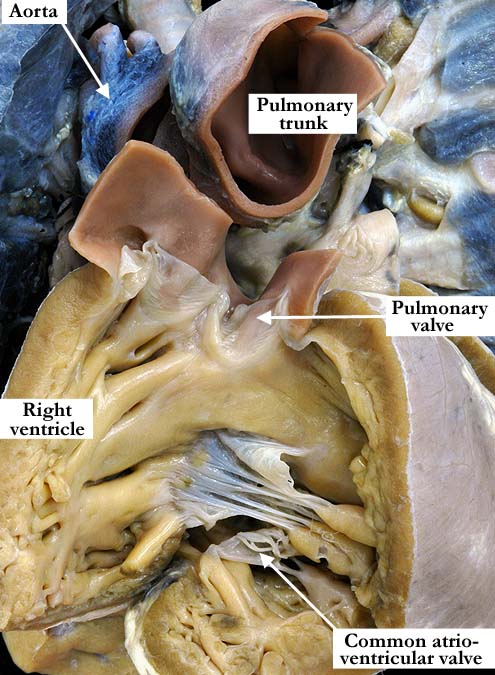
Modality: Anatomic specimen Orientation: Anterior view Description: This anterior view of the right ventricular outflow tract shows a markedly dilated pulmonary trunk. The pulmonary trunk was previously cut just above the valve. The common atrioventricular valve lies in the inlet. Contributor: Diane E. Spicer, BS Institution: The Congenital Heart Institute of Florida (CHIF) Image Label: A030105-99e Source of Image: Idriss Archive, Childrens Memorial Hospital, Chicago, IL Image Certification: pending AWG Rating: pending
|
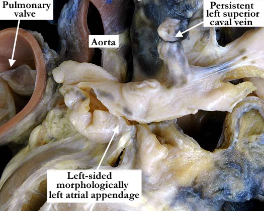
Modality: Anatomic specimen Orientation: Posterior lateral axis Description: This posterior lateral view shows a left-sided, morphologically left atrial appendage. The appendage is narrow and scalloped along the edges with a narrow attachment to the atrial vestibule. The left superior caval vein was ligated. Contributor: Diane E. Spicer, BS Institution: The Congenital Heart Institute of Florida (CHIF) Image Label: A030105-99f Source of Image: Idriss Archive, Childrens Memorial Hospital, Chicago, IL Image Certification: pending AWG Rating: pending
|
|||
|
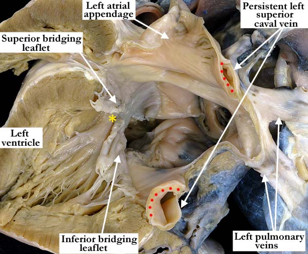
Modality: Anatomic specimen Orientation: Posterior view Description: The left-sided, morphologically left atrium and the left ventricle have been opened in a clam-shell fashion demonstrating the smooth atrial vestibule and pectinate muscles confined to the atrial appendage, these features consistent with left atrial morphology. The persistent left superior caval vein was bisected and clearly has a separate wall (red dots) from the left atrium as it extends over the posterior aspect of the left atrium to drain into the coronary sinus. The superior and inferior bridging leaflets of the common atrioventricular valve have a clear zone of apposition (astrisk) at the crest of the ventricular septum. Contributor: Diane E. Spicer, BS Institution: The Congenital Heart Institute of Florida (CHIF) Image Label: A030105-99g Source of Image: Idriss Archive, Childrens Memorial Hospital, Chicago, IL Image Certification: pending AWG Rating: pending
|
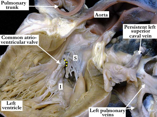
Modality: Anatomic specimen Orientation: Posterior view Description: The left ventricular free wall has been lifted away to show the left ventricular outflow tract and the aortic valve. The superior bridging leaflet (S) is in fibrous continuity with the aortic valve and the inferior bridging leaflet (I) lies against the interventricular septum. The zone of apposition between the two leaflets is clear and marked with yellow dots. Contributor: Diane E. Spicer, BS Institution: The Congenital Heart Institute of Florida (CHIF) Image Label: A030105-99h Source of Image: Idriss Archive, Childrens Memorial Hospital, Chicago, IL Image Certification: pending AWG Rating: pending
|
|||
AWG Page Certification: pending
|
Copyright ipccc-awg.net All Rights Reserved. Frontpage-Templates.org |