|
||||||||
|
|
|||||||||
|
|||||||||
|
IPCCC: 09.29.33, 01.04.04, 01.03.00, 02.03.01, 02.06.02, 07.02.00, 07.10.00,
01.05.01 |
|||
|
AEPC Derived Term: |
Interrupted aortic
arch between subclavian & common carotid arteries (type B) (09.29.33) |
||
|
EACTS-STS Derived Term: |
Interrupted aortic
arch (IAA), Type B (Interruption between the carotid and subclavian
arteries) (09.29.33) |
||
|
ICD-10 Derived Term: |
Other congenital malformations of aorta (Q25.4) Double inlet ventricle (Q20.4) |
||
|
Definition: pending
|
|
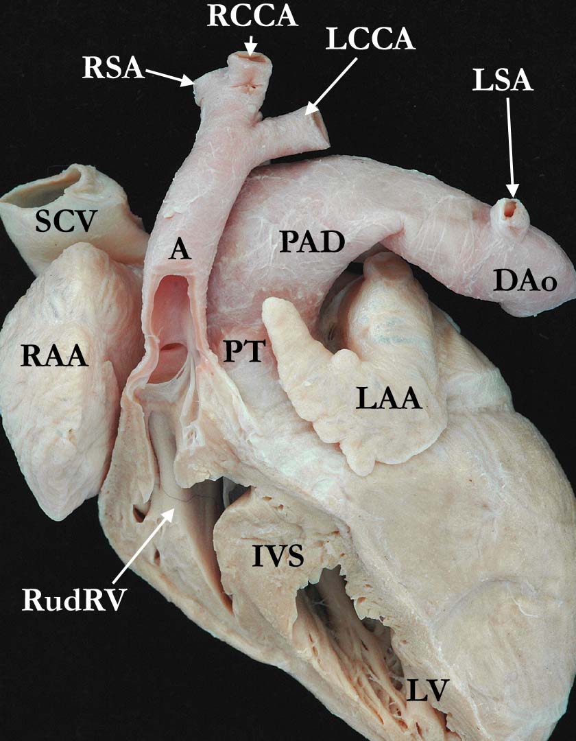
Modality: Anatomic specimen Orientation: Anterior view Description: This external view of the great arteries is viewed from the anterior, left side, illustrating an interrupted aortic arch. The aorta is interrupted between the left common carotid (LCCA) and the left subclavian (LSA) arteries. The left subclavian artery arises from the descending aorta (DAo) quite a distance from where the patent arterial duct (PAD) joins the aorta. The anterior aspect of the right and left ventricles have been obliquely resected in this heart with double inlet left ventricle (not shown), exposing the rudimentary right ventricle (RudRV), with the aorta (A) arising from it. The apical trabecular portion of the left ventricle (LV) and the interventricular septum (IVS) is seen. (LAA-left atrial appendage, PT-pulmonary trunk, RAA-right atrial appendage, RCCA-right common carotid, RSA-right subclavian artery, SCV-superior caval vein) Contributor: Diane Spicer, BS Institution: The Congenital Heart Institute of Florida (CHIF) Image Label: A010404-19a Source of Image: Van Mierop Archive, University of Florida, Gainesville, Florida Image Certification: 5 Nov 2011
AWG Rating:
|
||||
AWG Page Certification: 5 Nov 2011
|
Copyright ipccc-awg.net All Rights Reserved. Frontpage-Templates.org |