|
|||||
|
|
||||||
|
||||||
|
IPCCC: 09.29.13, 01.01.19, 07.09.00 |
|||
|
AEPC Derived Term: |
Aortic arch hypoplasia (tubular) between subclavian & common carotid arteries (09.29.13) Double outlet right ventricle: with non-committed VSD (01.01.19) Subaortic stenosis (07.09.00) |
||
|
EACTS-STS Derived Term: |
Aortic arch hypoplasia, Hypoplasia of aortic arch, Distal arch hypoplasia (distal to the carotid arteries and proximal to the subclavian artery) (09.29.13) VA connection =Double outlet VA connections, Double outlet RV, Remote VSD (Uncommitted VSD) (01.01.19) DORV-modifier, Aortic outflow tract obstruction, Subvalvar aortic obstruction present (07.09.00, 07.09.32) [Alternative classification: DORV-modifier, Aortic outflow tract obstruction, Transverse arch aortic obstruction present (09.28.20, 07.09.32)] |
||
|
Definition: pending |
|
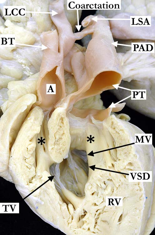
Modality: Anatomic specimen Orientation: Anterior view Description: The parietal wall of the right ventricle (RV) has been cut away to demonstrate the double outlet right ventricle. The aorta (A) and the pulmonary trunk (PT) are both supported by a complete muscular infundibulum (*). The subaortic infundibulum is long and narrow. Subaortic stenosis and coarctation of the aorta are anomalies that typically occur together. The great vessels branch normally from the aortic arch with tubular hypoplasia (coarctation) of the segment between the left common carotid (LCC) and left subclavian (LSA) arteries. The tricuspid valve (TV) guards the inlet of the right ventricle and a small portion of the mitral valve (MV) can be seen through the uncommitted ventricular septal defect (VSD). (BT-brachiocephalic trunk, PAD-patent arterial duct, PV-pulmonic valve) Contributor: Diane Spicer, BS Institution: The Congenital Heart Institute of Florida (CHIF) Image Label: A092913-63a Source of Image: Idriss Archive, Childrens Memorial Hospital, Chicago, IL Image Certification: pending AWG Rating: pending
|
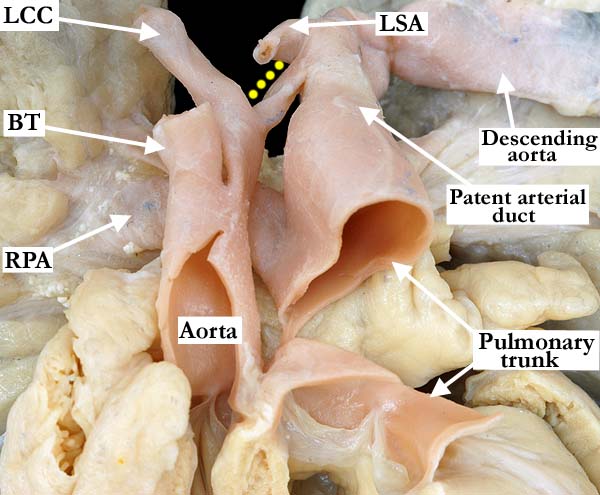
Modality: Anatomic specimen Orientation: Anterior view Description: This anterior, anatomic view of the great vessels as they exit the base of the heart shows the aorta to the right of the pulmonary artery (Note: The pulmonary trunk has been nearly transected). There is tubular hypoplasia of the segment of aortic arch (yellow dots) between the left common carotid (LCC) and left subclavian (LSA) arteries. The pulmonary trunk is dilated and the patent arterial duct extends directly into the descending aorta. (Note: Just distal to where the arterial duct connects to the descending aorta, there is an artifactual constriction of the aorta). (RPA-right pulmonary artery, BT-brachiocephalic trunk) Contributor: Diane Spicer, BS Institution: The Congenital Heart Institute of Florida (CHIF) Image Label: A092913-63b Source of Image: Idriss Archive, Childrens Memorial Hospital, Chicago, IL Image Certification: pending AWG Rating: pending |
|||
AWG Page Certification: pending
|
Copyright ipccc-awg.net All Rights Reserved. Frontpage-Templates.org |