|
||||||||
|
|
|||||||||
|
|||||||||
|
IPCCC: 09.29.22, 09.29.12, 01.04.04, 01.03.00, 02.03.01,
02.06.02, 07.02.00, 07.10.00, 01.05.01, 01.06.01, 07.14.01 |
|||
|
AEPC Derived Term: |
Aortic arch atresia:
fibrous cord distal to subclavian artery (type A) (09.29.22) Two AV valves in double inlet ventricle (01.06.01) Restrictive VSD (07.14.01) |
||
|
EACTS-STS Derived Term: |
Aortic pathology,
Abnormality involving aortic arch, Aortic arch atresia, Fibrous cord,
Atresia distal to subclavian artery (type A) (09.29.22) |
||
|
ICD-10 Derived Term: |
Other congenital malformations of aorta (Q25.4) Double inlet ventricle (Q20.4) |
||
|
Definition: pending
|
|
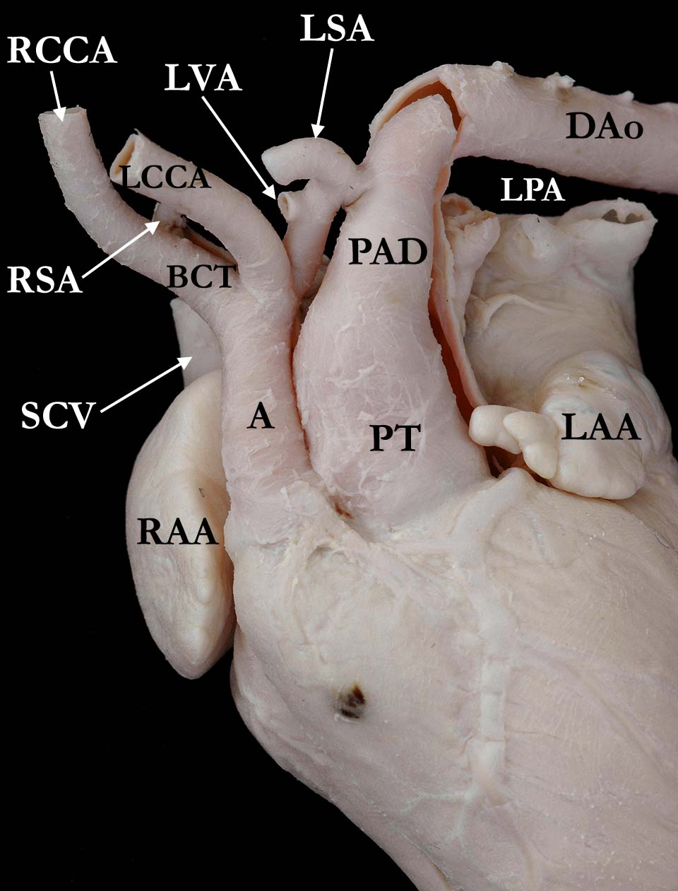
Modality: Anatomic specimen Orientation: Right anterior view Description: This view shows the outward appearance of a heart with aortic arch atresia and transposition of the great arteries with double inlet left ventricle (ventricular inlet not shown). The ascending aorta (A) is relatively small and is located to the right of the pulmonary trunk (PT). The aortic isthmus is hypoplastic between the left common carotid (LCCA) and the patent arterial duct (PAD). This severely stenotic segment is patent up to and including the left subclavian artery (LSA). The aorta then becomes an atretic, fibrous segment where it attaches to the arterial duct (seen on further dissection). Note the left subclavian artery is smaller than the other brachiocephalic vessels. (BCT-brachiocephalic trunk, DAo-descending aorta, LAA-left atrial appendage, LVA-vertebral artery, PT-pulmonary trunk, RAA-right atrial appendage, RCCA-right common carotid, RSA-right subclavian artery, SCV-superior caval vein) Contributor: Diane Spicer, BS Institution: The Congenital Heart Institute of Florida (CHIF) Image Label: A010404-18a Source of Image: Van Mierop Archive, University of Florida, Gainesville, Florida Image Certification: 5 Nov 2011
AWG Rating:
|
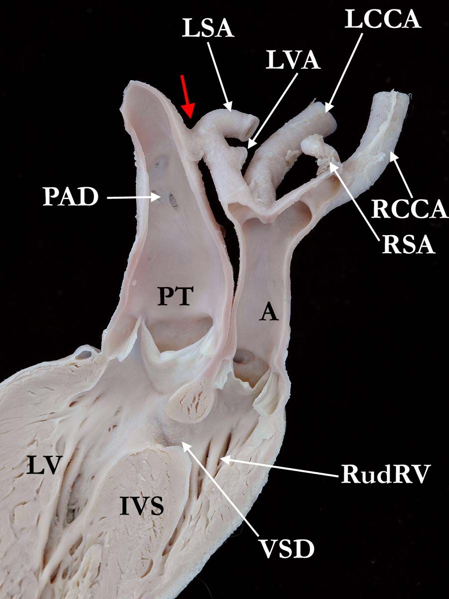
Modality: Anatomic specimen Orientation: Long axis view
Description: This view is a long axis view
of the great arteries as they exit the ventricular mass. It shows the
luminal surface of the vessels and illustrates the atretic portion of the
aorta (red arrow) as well as the severe coarctation of the isthmus. Note the
relatively small ascending aorta (A) arising from the rudimentary right
ventricle (RudRV)
along with the ventricular septal defect and the pulmonary
trunk arising from the left ventricle (LV).
This view shows that the
ventricular septal defect (VSD) is smaller than the aortic orifice;
therefore, likely restrictive. (IVS-interventricular septum, LCCA-left common
carotid artery,
LVA-vertebral artery, PAD-patent arterial duct, PT-pulmonary
trunk,
RCCA-right common carotid artery,
RSA-right subclavian artery,
SCV-superior
caval vein, VA-vertebral artery). Institution: The Congenital Heart Institute of Florida (CHIF) Image Label: A010404-18b Source of Image: Van Mierop Archive, University of Florida, Gainesville, Florida Image Certification: 5 Nov 2011
AWG Rating:
|
|||
AWG Page Certification: 5 Nov 2011
|
Copyright ipccc-awg.net All Rights Reserved. Frontpage-Templates.org |