|
||||||||
|
|
|||||||||
|
|||||||||
|
IPCCC:
06.07.05, 06.06.09 |
|||
|
AEPC Derived Term: |
Atrioventricular septal defect (AVSD) with ventricular imbalance: dominant right ventricle, hypoplastic left ventricle (06.07.05) Atrioventricular septal defect (AVSD): atrial & ventricular components with common atrioventricular orifice (complete) (06.06.09) |
||
|
EACTS-STS Derived Term: |
AVC (AVSD), Complete (CAVSD), Unbalanced, Small LV (06.07.05, 06.06.09) |
||
|
ICD10 Derived Term: |
Atrioventricular septal defect (Q21.2) |
||
|
Definition: pending
|
|
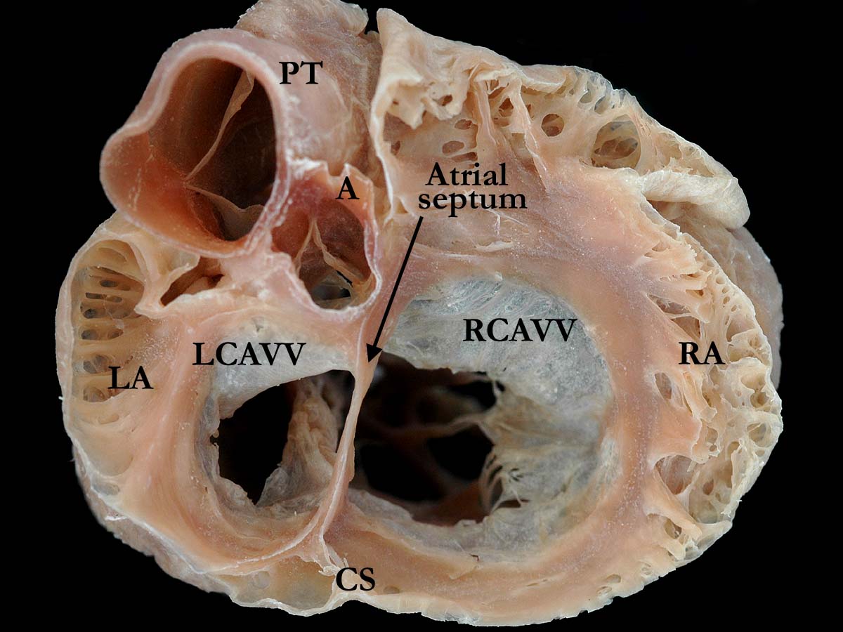
Modality: Anatomic specimen Orientation: Short axis view Description: This short axis view of the base of the heart demonstrates an unbalanced atrioventricular septal defect. The right portion of this common atrioventricular valve is dominant. The right atrium (RA) is larger and dilated when compared to the hypoplastic left atrium (LA). A strand of atrial septal tissue is easily visualized extending from the crux of the heart toward the anterior superior aspect of the heart. Note the unwedged position of the aorta (A). There are concordant atrioventricular and ventriculoarterial connections. (CS-coronary sinus, LCAVV-left component of the common atrioventricular valve, RCAVV-right component of the common atrioventricular valve, PT-pulmonary trunk). Contributor: Diane E. Spicer, BS Institution: The Congenital Heart Institute of Florida (CHIF) Image Label: A060705-15a Source of Image: Van Mierop Archive, University of Florida, Gainesville, Florida Image Certification: 2 July 2011, (rediscussed 3 March 2012)
AWG Rating:
|
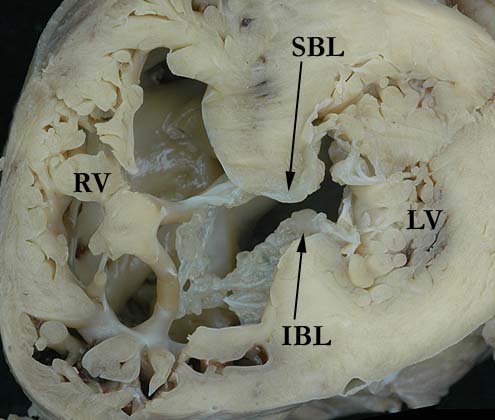
Modality: Anatomic specimen Orientation: Short axis view Description: This short axis view from the apex of the heart demonstrates an unbalanced common atrioventricular valve where the right side is dominant. The superior (SBL) and inferior (IBL) bridging leaflets of the common valve are easily visualized. Contributor: Diane E. Spicer, BS Institution: The Congenital Heart Institute of Florida (CHIF) Image Label: A060705-15b Source of Image: Van Mierop Archive, University of Florida, Gainesville, Florida Image Certification: 2 July 2011, (rediscussed 3 March 2012)
AWG Rating:
|
|||
AWG Page Certification: 2 July 2011 (rediscussed 3 March 2012)
|
Copyright ipccc-awg.net All Rights Reserved. Frontpage-Templates.org |