|
||||||||
|
|
|||||||||
|
|||||||||
|
IPCCC: 06.06.01 |
|||
|
AEPC Derived Term: |
AVSD: isolated atrial component (primum ASD)(partial) (06.06.01) |
||
|
EACTS-STS Derived Term: |
ASD, Primum (06.06.01) |
||
|
ICD10 Derived Term: |
Atrioventricular septal defect (Q21.2) | ||
|
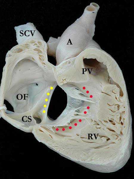
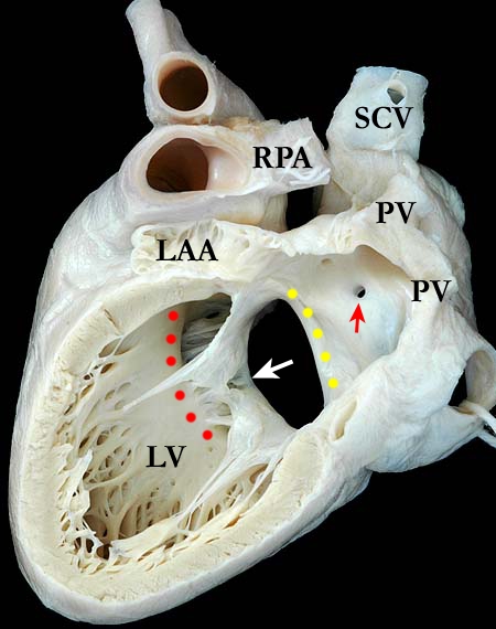
Definition: A congenital cardiac malformation that is a variant of an atrioventricular septal defect (atrioventricular canal) with an interatrial communication just above the atrioventricular valve, no interventricular communication just below the atrioventricular valve, separate right and left atrioventricular valvar orifices, and varying degrees of malformation of the left sided component of the common atrioventricular valve. The bridging leaflets of the common atrioventricular valve are bound down to the crest of the scooped out ventricular septum so that the potential for shunting through the atrioventricular septal defect is possible only at the atrial level and not at the ventricular level.
Discussion: The subcostal long axis views of both the right (A060601-13a) and left (A060601-13b) sides of the heart show a common atrioventricular junction with separate valvar orifices, this also known as an ostium primum defect. The superior and inferior bridging leaflets are attached by a connecting tongue of valve tissue, creating the separate right and left valvar orifices. The valve tissue is adherent along the entire length of the ventricular septum. There are concordant atrioventricular and ventriculo-arterial connections. The characteristic scooping out (red dots) of the ventricular septum is easily appreciated and shunting would occur entirely at the atrial level. The lower edge of the atrial septum is illustrated with yellow dots. The upper portion (oval fossa or fossa ovalis) of the atrial septum is virtually intact with a tiny probe patent defect. (A-aorta, CS-coronary sinus, LAA-left atrial appendage, LV-left ventricle, OF-oval fossa, PV-pulmonary valve, PVV-pulmonary vein, RPA-right pulmonary artery, RV-right ventricle, SCV-superior caval vein) |
|
Modality: Anatomic specimen Orientation: Right subcostal long axis Description: In this view, the common atrioventricular valve is clearly separated from the pulmonary valve (PV) by a muscular infundibulum. Contributor: Diane Spicer, BS Institution: The Congenital Heart Institute of Florida (CHIF) Image Label: A060601-13a Source of Image: Van Mierop Archive, University of Florida, Gainesville, Florida Image Certification: 5 August 2011
AWG Rating:
|
Modality: Anatomic specimen Orientation: Left subcostal long axis Description: In this instance the zone of apposition (white arrow) is distant from the ventricular septal crest where as in most other specimens with the same defect, the zone of apposition lies adjacent to the ventricular septal crest. The upper portion of the atrial septum is well formed with a tiny septal defect (red arrow). Contributor: Diane Spicer, BS Institution: The Congenital Heart Institute of Florida (CHIF) Image Label: A060601-13b Source of Image: Van Mierop Archive, University of Florida, Gainesville, Florida Image Certification: 5 August 2011
AWG Rating:
|
|||
AWG Page Certification: 5 August 2011
|
Copyright ipccc-awg.net All Rights Reserved. Frontpage-Templates.org |