|
|||||
|
|
||||||
|
||||||
|
IPCCC: 06.06.09, 05.04.02 |
|||
|
AEPC Derived Term: |
AVSD: atrial &
ventricular components with common AV orifice (complete) (06.06.09) |
||
|
EACTS-STS Derived Term: |
AVC (AVSD), Complete (CAVSD) (06.06.09) ASD, Secundum (05.04.02) |
||
|
Definition: pending
|
|
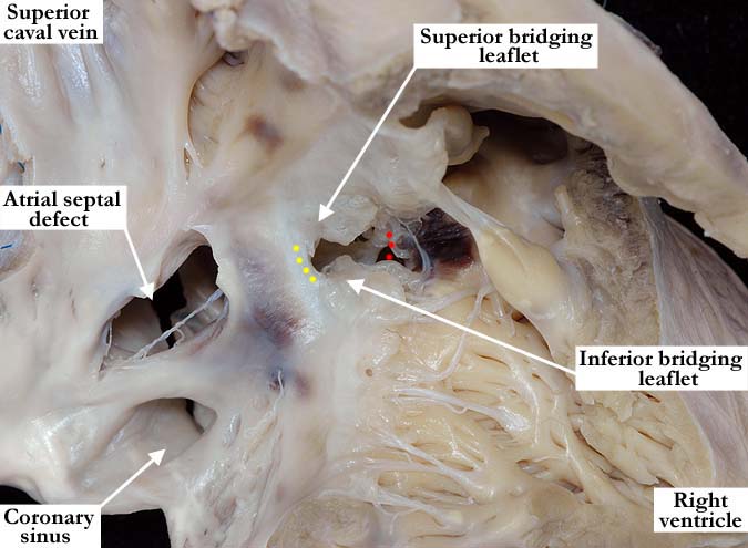
Modality: Anatomic specimen Orientation: Right ventricular view Description: In this anatomic view, the free wall of the right ventricle has been lifted away to show the septal surfaces of the right atrium and ventricle. The upper portion of the atrial septum is well formed with a large secundum, atrial septal defect. The coronary sinus is in its usual position. There is a common atrioventricular valve with a restrictive, atrioventricular septal defect. The superior and inferior bridging leaflets are attached to the atrial septum (yellow dots), allowing for shunting to take place only at the ventricular level. (red dots-crest of the ventricular septum) Contributor: Diane E. Spicer, BS Institution: The Congenital Heart Institute of Florida (CHIF) Image Label: A060609-87a Image Source: Medical University of South Carolina Image Certification: pending AWG Rating: pending
|
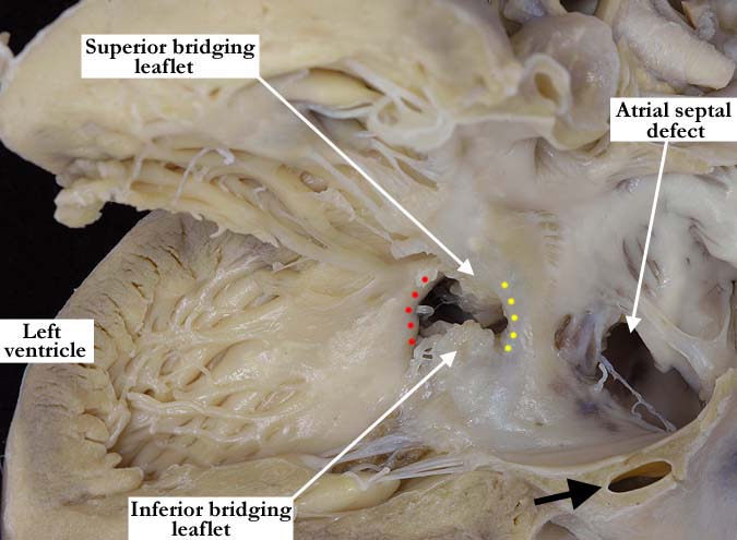
Modality: Anatomic specimen Orientation: Left ventricular view Description: This anatomic view of the left atrial and ventricular septum of the heart demonstrates the left side of this restrictive, atrioventricular septal defect. The superior and inferior bridging leaflets of the common atrioventricular valve are attached to the atrial portion (yellow dots) of the defect. This effectively allows for shunting to occur only at the ventricular level. There is a large secundum atrial septal defect and the vein of the left atrium (black arrow) lies within the inferior wall of the left atrium as it extends to its normal position in the left atrioventricular groove. (red dots-crest of the ventricular septum) Contributor: Diane E. Spicer, BS Institution: The Congenital Heart Institute of Florida (CHIF) Image Label: A060609-87b Image Source: Medical University of South Carolina Image Certification: pending AWG Rating: pending
|
|||
AWG Page Certification: pending
|
Copyright ipccc-awg.net All Rights Reserved. Frontpage-Templates.org |