|
|||||
|
|
||||||
|
||||||
|
IPCCC:
06.07.27, 06.07.04, 06.06.09, 09.29.01, 09.29.12
|
|||
|
AEPC Derived Term: |
AVSD with balanced ventricles (06.07.27) AVSD ventricular component under superior bridging leaflet: free floating & chords to papillary muscle at right ventricular apex-free wall (Rastelli C) (06.07.04) AVSD: atrial & ventricular components with common AV orifice (complete) (06.06.09) Aortic coarctation (09.29.01) Aortic arch hypoplasia (tubular) distal to subclavian artery (isthmal) (09.29.12) |
||
|
EACTS-STS Derived Term: |
AVC (AVSD), Complete (CAVSD), Balanced, Rastelli type C (06.07.27, 06.07.04, 06.06.09) Coarctation of the Aorta (CoAo)-modifier, With isthmus hypolasia (distal to the subclavian artery), (09.29.01, 09.29.12) |
||
|
Definition: pending
|
|
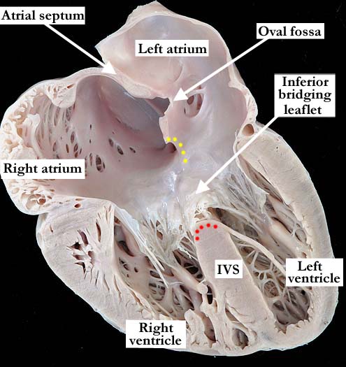
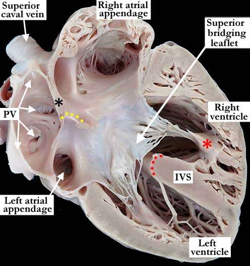
Modality: Anatomic specimen Orientation: Simulated four chamber echocardiographic view, inferior portion of heart (left image), anterior portion of heart (right image) Description: These images demonstrate simulated four chamber echocardiographic views of a heart with a complete atrioventricular septal defect and concordant atrioventricular connections. The upper portion of the atrial septum is well formed with an atrial septal defect at the oval fossa. The yellow dots mark the atrial border of the atrioventricular septal defect with the red dots on the crest of the interventricular septum (IVS), marking the ventricular component of the defect. The inferior bridging leaflet in the left image is bound to the crest of the interventricular septum by multiple tendinous cords, while the superior bridging leaflet in the right image is free floating. The superior bridging leaflet is attached to an anterior papillary muscle and has a larger commitment to the right ventricle. Under the Rastelli Classification, this is type 3. Contributor: Diane E. Spicer, BS Institution: The Congenital Heart Institute of Florida (CHIF) Image Label: A060727-84a (left image); A060727-84b (right image) Image Source: Van Mierop Archive, University of Florida Image Certification: pending AWG Rating: pending
|
|
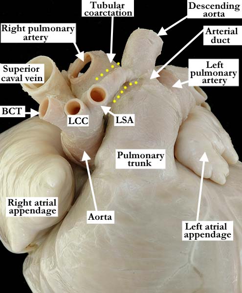
Modality: Anatomic specimen Orientation: Antero-superior Description: This antero-superior anatomic view demonstrates normally related great arteries, the aorta somewhat smaller than the pulmonary trunk. The brachiocephalic trunk (BCT), the left common carotid (LCC) and the left subclavian artery (LSA) branch from the arch in the usual fashion. Distal to the left subclavian artery there is a preductal, tubular coarctation (yellow dots). This type of coarctation is known as tubular hypoplasia and is characterized by the uniform narrowing of the aortic arch. Contributor: Diane E. Spicer, BS Institution: The Congenital Heart Institute of Florida (CHIF) Image Label: A060727-84c Image Source: Van Mierop Archive, University of Florida Image Certification: pending AWG Rating: pending
|
||||
AWG Page Certification: pending
|
Copyright ipccc-awg.net All Rights Reserved. Frontpage-Templates.org |