|
||||||||
|
|
|||||||||
|
|||||||||
|
IPCCC: 06.06.09, 06.05.03, 03.01.04 or 01.03.02
|
|||
|
AEPC Derived Term: |
AVSD: atrial & ventricular
components with common AV orifice (complete) (06.06.09) |
||
|
EACTS-STS Derived Term: |
AVC (AVSD), Complete (CAVSD), AVC (AVSD)-modifier, AVSD AV valvar
abnormality (06.06.09) Common AV valvar dysplasia (mucoid thickening) (06.05.03) Atrial appendage isomerism, Right (03.01.04) |
||
|
ICD10 Derived Term: |
Atrioventricular septal defect (Q21.2) Other specified congenital malformations of heart (Q24.8) Isomerism of atrial appendages (Q20.6) |
||
|
Definition: pending
|
|
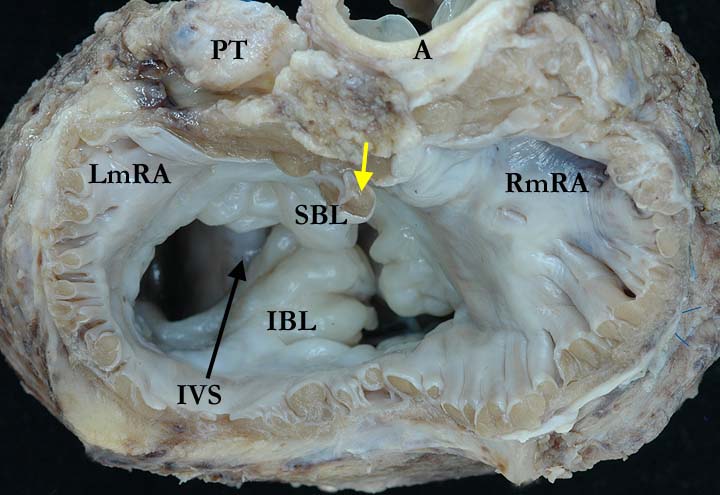
Modality: Anatomic specimen Orientation: Short axis view Description: This short axis view, from the base of the heart, shows the characteristic features of a common atrioventricular junction. The aorta has been sprung from its usual wedged position between the two atrioventricular valves and there is a common atrioventricular valve. The valve is very dysplastic and appears as though the superior (SBL) and inferior (IBL) bridging leaflets are fused. In reality, they appose one another and are not fused (see image A060609-11b), floating freely over the interventricular septum (IVS). Note the pectinate muscles extending around the entire common atrioventricular junction in this heart with right isomerism. the yellow arrow marks the remnant of the atrial septum. (LmRA-left sided, morphologically right atrium, RmRA-right sided, morphologically right atrium) Contributor: Diane E. Spicer, BS Institution: The Congenital Heart Institute of Florida (CHIF) Image Label: A060609-11a Source of Image: The Congenital Heart Institute of Florida (CHIF) Image Certification: 7 May 2011
AWG Rating:
|
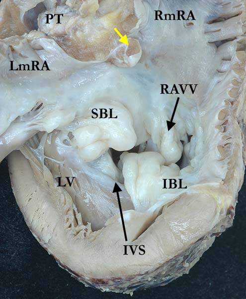
Modality: Anatomic specimen Orientation: Left ventricular view Description: This view shows the highly dysplastic, common atrioventricular valve, the superior (SBL) and inferior (IBL) bridging leaflets, straddling the interventricular septum. The right portion of the common atrioventricular valve (RAVV) is easily appreciated. The yellow arrow marks the remnant of the atrial septum. ( LmRA-left sided, morphologically right atrium, PT-pulmonary trunk, RmRA-right sided, morphologically right atrium) Contributor: Diane E. Spicer, BS Institution: The Congenital Heart Institute of Florida (CHIF) Image Label: A060609-11b Source of Image: The Congenital Heart Institute of Florida (CHIF) Image Certification: 7 May 2011
AWG Rating:
|
|||
AWG Page Certification: 7 May 2011
|
Copyright ipccc-awg.net All Rights Reserved. Frontpage-Templates.org |