|
||||||||
|
|
|||||||||
|
|||||||||
|
IPCCC: 05.05.02, 4.06.23, Q1.01.52, 04.01.03, 04.01.26 |
|||
|
AEPC Derived Term: |
Sinus venosus defect
(ASD) with overriding inferior caval vein (IVC) (inferior defect)(05.05.02) |
||
|
EACTS-STS Derived Term: |
ASD, Sinus venosus, IVC type (05.05.02) |
||
|
ICD10 Derived Term: |
Atrial septal defect (Q21.1) Other congenital malformations of
great veins (Q26.8) |
||
|
Definition: pending
|
|
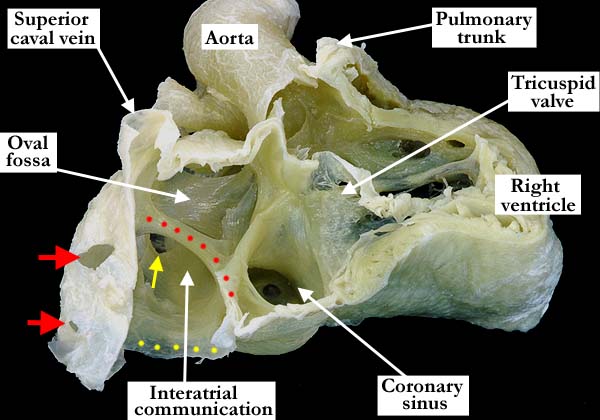
Modality: Anatomic specimen Orientation: Right anterior oblique view Description: In this view, the heart has been tilted slightly upwards. The free wall of the right atrium and ventricle has been removed. The superior caval vein drains to the right atrium in the usual fashion. There is a persistent left superior caval vein which drains to the dilated coronary sinus. The oval fossa is patent. Along the inferior border of the oval fossa, the red dots mark the roof of a sinus venosus defect or interatrial communication associated with the inferior caval vein. The inferior border of the inferior caval vein is identified by yellow dots. The right pulmonary veins (red arrows) drain in an anomalous fashion to the inferior caval vein. One left pulmonary vein (yellow arrow) is visualized through the defect, all of the left pulmonary veins draining in the appropriate fashion to the left atrium. Contributor: Diane E. Spicer, BS Institution: The Congenital Heart Institute of Florida (CHIF) Image Label: A050502-7a Image Source: Van Mierop Archive, University of Florida Image Certification: 7 May 2011
AWG Rating:
|
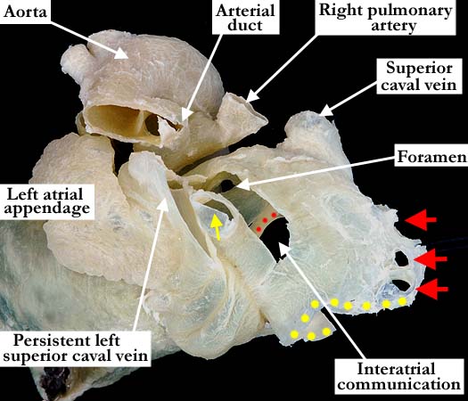
Modality: Anatomic specimen Orientation: Posterior view Description: This posterior view of the left atrium demonstrates the persistent left superior caval vein that lies over the posterior surface of the left atrium, between the left atrial appendage and the left pulmonary venous component (yellow arrow). Through a slit-like opening in the left atrium a portion of the foramen and the superior rim (red dots) of the sinus venosus defect (interatrial communication) are seen. The anterior lateral aspect of the inferior caval vein has been stretched out to view the anomalous right pulmonary venous return (red arrows). The inferior border of the inferior caval vein is marked with yellow dots. Contributor: Diane E. Spicer, BS Institution: The Congenital Heart Institute of Florida (CHIF) Image Label: A050502-7b Image Source: Van Mierop Archive, University of Florida Image Certification: 7 May 2011
AWG Rating:
|
|||
AWG Page Certification: 7 May 2011
|
Copyright ipccc-awg.net All Rights Reserved. Frontpage-Templates.org |