|
|||||
|
|
||||||
|
||||||
|
IPCCC: 05.04.02 |
|||
|
AEPC Derived Term: |
Atrial septal defect (ASD) within oval fossa (secundum) (05.04.02) | ||
|
EACTS-STS Derived Term: |
ASD, Secundum (05.04.02) | ||
|
Definition: pending |
|
Modality: Anatomic specimen Orientation: Right atrium and right ventricle opened and viewed in anatomic position Description: The superior (SCV) and inferior (ICV) caval veins enter the morphologic right atrium in the usual fashion. There is a large secundum atrial septal defect (ASD). The tricuspid valve (TV) guards the inlet and the coronary sinus (CS) is in its usual position (covered by the Thebesian valve). Contributor: Diane Spicer, BS Institution: Congenital Heart Institute of Florida Image Label: A050402-56a Image Certification: 24 July 2010
AWG Rating:
|
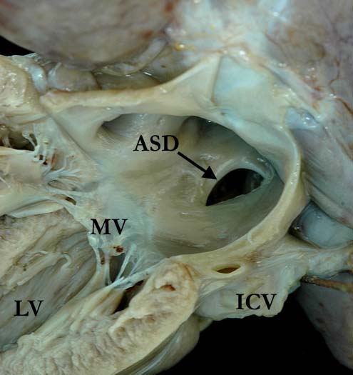
Modality: Anatomic specimen Orientation: Left atrium and left ventricle opened and viewed in anatomic position Description: The left atrial aspect of the secundum atrial septal defect (ASD) seen in image A050402-56a. The mitral valve (MV) guards the inlet to the morphologic left ventricle (LV). (ICV - inferior caval vein) Contributor: Diane Spicer, BS Institution: Congenital Heart Institute of Florida Image Label: A050402-56b Image Certification: 24 July 2010
AWG Rating:
|
|||
|
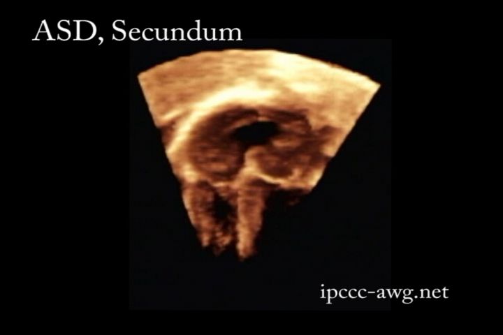
Modality: Transthoracic 3D echocardiogram Orientation: Apical 4-chamber Description: This image shows a secundum atrial septal defect (ASD). Contributor: Stan Timofeev, MS, RDCS, RVS Institution: Congenital Heart Institute of Florida Image Label: E050402-56c Image Certification: 24 July 2010
AWG Rating:
|
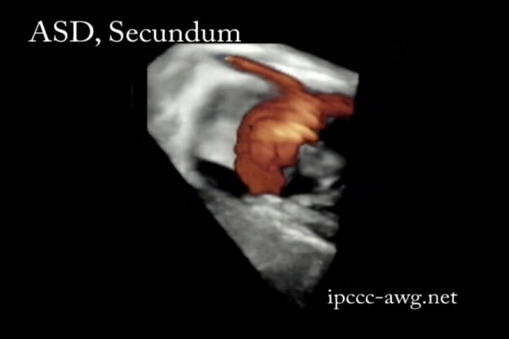
Modality: Transthoracic 3D echocardiogram Orientation: Apical 4-chamber Description: This image shows a secundum atrial septal defect (ASD) and flow (color doppler) across the atrial septal defect. Note the pulmonary veins draining into the left atrium. There is some element of tricuspid regurgitation present as well. Contributor: Stan Timofeev, MS, RDCS, RVS Institution: Congenital Heart Institute of Florida Image Label: E050402-56d Image Certification: 24 July 2010
AWG Rating:
|
AWG Page Certification: 24 July 2010
|
Copyright ipccc-awg.net All Rights Reserved. Frontpage-Templates.org |