|
||||||||
|
|
|||||||||
|
|||||||||
|
IPCCC: 09.04.01, 09.29.22, 09.41.02 |
|||
|
AEPC Derived Term: |
Aorto-pulmonary window (09.04.01) Aortic arch atresia: fibrous cord distal to subclavian artery (type A) (09.29.22) Anomalous origin of right coronary artery from pulmonary artery (ARCAPA) (09.41.02) |
||
|
EACTS-STS Derived Term: |
AP window and Interrupted aortic arch (09.04.01, 09.29.31) Aortic pathology, Abnormality involving aortic arch, Aortic arch atresia, Fibrous cord, Atresia distal to subclavian artery (type A) (09.29.22) Coronary anomaly, APOC (Anomalous pulmonary origin of coronary), ARCAPA (09.41.02) |
||
|
ICD10 Derived Term: |
Aortopulmonary septal defect (Q21.4) Atresia of aorta (Q25.2) |
||
|
Definition: pending |
|
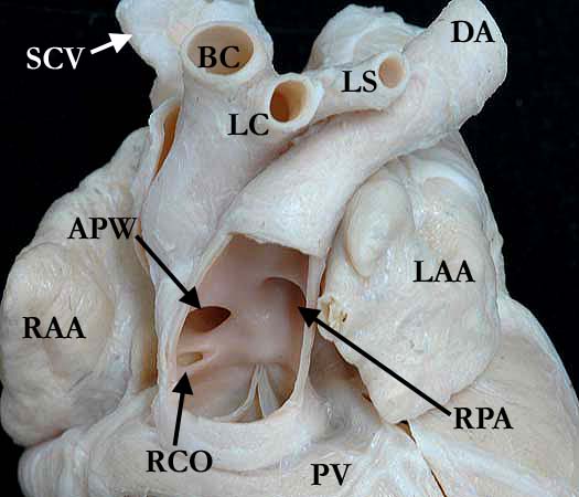
Modality: Anatomic specimen Orientation: Anterior-superior view Description: The pulmonary trunk has been resected anteriorly to show the aorto-pulmonary window (APW). The defect is seen in the right lateral wall of the pulmonary trunk with the right coronary orifice (RCO) just inferior to the defect. There is a separate aortic valve not shown in this image. The right pulmonary artery (RPA) origin is just superior and to the left of the aorto-pulmonary window (APW). The left pulmonary artery origin is distal and not shown in this image. The aortic arch is interrupted distal to the left subclavian artery (LS) and the arterial duct is widely patent. (SCV-superior caval vein, RAA-right atrial appendage, LAA-left atrial appendage, BC-brachiocephalic trunk, LC-left common carotid artery, DA-descending aorta, PV-pulmonary valve) Contributor: Diane Spicer, BS Institution: The Congenital Heart Institute of Florida (CHIF) Image Label: A090401-3a Source of Image: Van Mierop Archive, University of Florida, Gainesville, FL Image Certification: 5 February 2011
AWG Rating:
|
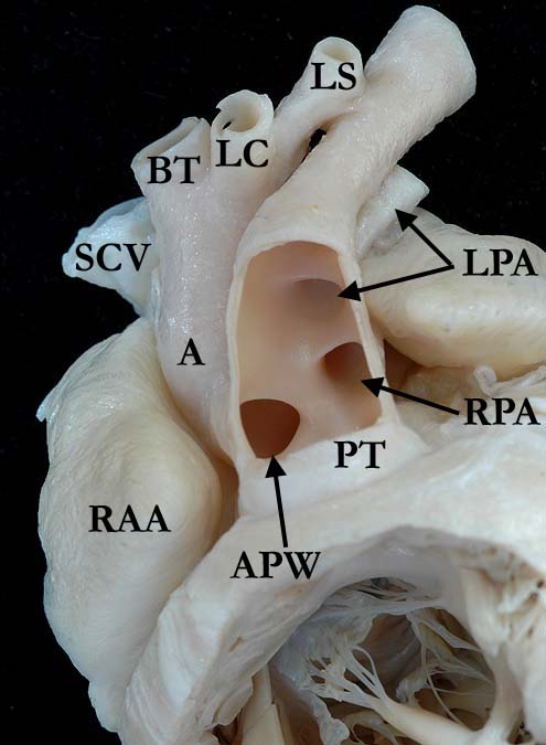
Modality: Anatomic specimen Orientation: Anterior view Description: This image is similar to A090401-3a but angled slightly inferior to show both the right and left pulmonary orifices as they exit the pulmonary trunk. The pulmonary trunk has been resected anteriorly to show the aorto-pulmonary window (APW). The defect is seen in the right lateral wall of the pulmonary trunk with the right coronary orifice (RCO) just inferior to the defect. There is a separate aortic valve not shown in this image. The right pulmonary artery (RPA) origin is just superior and to the left of the aorto-pulmonary window (APW). The left pulmonary artery origin is distal and not shown in this image. The aortic arch is interrupted distal to the left subclavian artery (LS) and the arterial duct is widely patent. (SCV-superior caval vein, RAA-right atrial appendage, LAA-left atrial appendage, BC-brachiocephalic trunk, LC-left common carotid, DA-descending aorta, PV-pulmonary valve) Contributor: Diane Spicer, BS Institution: The Congenital Heart Institute of Florida (CHIF) Image Label: A090401-3b Source of Image: Van Mierop Archive, University of Florida, Gainesville, FL Image Certification: 5 February 2011
AWG Rating:
|
|||
|
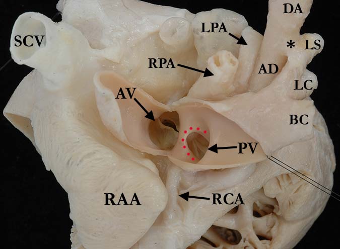
Modality: Anatomic specimen Orientation: Superior view Description: This view of the base of the heart from the opened aorta shows the aorto-pulmonary window (red dots) along with the separate aortic (AV) and pulmonary (PV) valves. The aortic valve is bicuspid. The aortic arch is directed leftward demonstrating the normal branching of the brachiocephalic trunk (BC), left common carotid (LC) and left subclavian (LS) arteries from the posterior aspect. An asterisk (*) denotes the fibrous cord of the atretic arch (the location of luminal aortic interruption). The right coronary artery (RCA) extends from the pulmonary trunk to the right atrioventricular groove. (RPA-right pulmonary artery, LPA-left pulmonary artery, AD-arterial duct, DA-descending aorta) Contributor: Diane Spicer, BS Institution: The Congenital Heart Institute of Florida (CHIF) Image Label: A090401-3c Source of Image: Van Mierop Archive, University of Florida, Gainesville, FL Image Certification: 5 February 2011
AWG Rating:
|
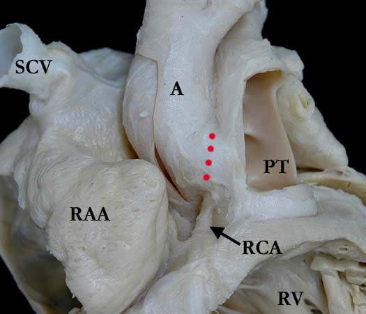
Modality: Anatomic specimen Orientation: Right anterior oblique view Description: This external view of the aorto-pulmonary window (red dots) shows the confluence between the aorta (A) and pulmonary trunk (PT) where the defect occurs. The right coronary artery (RCA) exits the pulmonary trunk just inferior to the defect and extends along the right atrioventricular groove in the usual fashion. This heart does have a separate aortic valve and pulmonary valve, although these spearate valves cannot be visialized in this image. (SCV-superior caval vein, RAA-right atrial appendage, RV-right ventricle) Contributor: Diane Spicer, BS Institution: The Congenital Heart Institute of Florida (CHIF) Image Label: A090401-3d Source of Image: Van Mierop Archive, University of Florida, Gainesville, FL Image Certification: 5 February 2011
AWG Rating:
|
|||
AWG Page Certification: 5 February 2011
|
Copyright ipccc-awg.net All Rights Reserved. Frontpage-Templates.org |